4U8H | pdb_00004u8h

Crystal Structure of Mammalian Period-Cryptochrome Complex


Experimental Data Snapshot

  • Method: X-RAY DIFFRACTION
  • Resolution: 2.80 Å
  • R-Value Free: 
    0.278 (Depositor), 0.279 (DCC) 
  • R-Value Work: 
    0.205 (Depositor), 0.213 (DCC) 
  • R-Value Observed: 
    0.209 (Depositor) 

Starting Model: experimental
View more details

wwPDB Validation 3D Report Full Report

Validation slider image for 4U8H

This is version 1.4 of the entry. See complete history

Literature

Molecular assembly of the period-cryptochrome circadian transcriptional repressor complex.

Nangle, S.N.Rosensweig, C.Koike, N.Tei, H.Takahashi, J.S.Green, C.B.Zheng, N.

(2014) Elife 3: e03674-e03674

  • DOI: https://doi.org/10.7554/eLife.03674
  • Primary Citation Related Structures: 
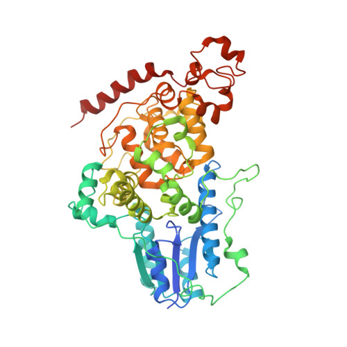
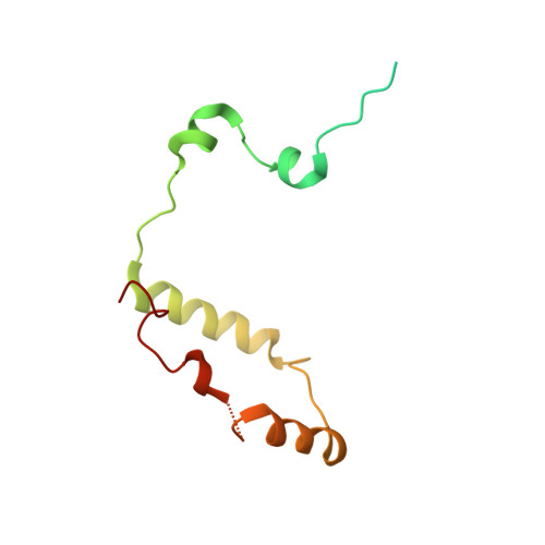
    4U8H

  • PubMed Abstract: 

    The mammalian circadian clock is driven by a transcriptional-translational feedback loop, which produces robust 24-hr rhythms. Proper oscillation of the clock depends on the complex formation and periodic turnover of the Period and Cryptochrome proteins, which together inhibit their own transcriptional activator complex, CLOCK-BMAL1. We determined the crystal structure of the CRY-binding domain (CBD) of PER2 in complex with CRY2 at 2.8 Å resolution. PER2-CBD adopts a highly extended conformation, embracing CRY2 with a sinuous binding mode. Its N-terminal end tucks into CRY adjacent to a large pocket critical for CLOCK-BMAL1 binding, while its C-terminal half flanks the CRY2 C-terminal helix and sterically hinders the recognition of CRY2 by the FBXL3 ubiquitin ligase. Unexpectedly, a strictly conserved intermolecular zinc finger, whose integrity is important for clock rhythmicity, further stabilizes the complex. Our structure-guided analyses show that these interspersed CRY-interacting regions represent multiple functional modules of PERs at the CRY-binding interface.


  • Organizational Affiliation
    • Department of Pharmacology, University of Washington, Seattle, United States.

Macromolecule Content 

  • Total Structure Weight: 144.74 kDa 
  • Atom Count: 9,342 
  • Modeled Residue Count: 1,138 
  • Deposited Residue Count: 1,262 
  • Unique protein chains: 2

Macromolecules

Find similar proteins by:|  3D Structure
Entity ID: 1
MoleculeChains  Sequence LengthOrganismDetailsImage
Cryptochrome-2A,
B [auth C]
510Mus musculusMutation(s): 0 
Gene Names: Cry2Kiaa0658
UniProt
Find proteins for Q9R194 (Mus musculus)
Explore Q9R194 
Go to UniProtKB:  Q9R194
Entity Groups
Sequence Clusters30% Identity50% Identity70% Identity90% Identity95% Identity100% Identity
UniProt GroupQ9R194
Sequence Annotations
Expand
Reference Sequence
Find similar proteins by:|  3D Structure
Entity ID: 2
MoleculeChains  Sequence LengthOrganismDetailsImage
Period circadian protein homolog 2C [auth B],
D
121Mus musculusMutation(s): 0 
Gene Names: Per2
UniProt & NIH Common Fund Data Resources
Find proteins for O54943 (Mus musculus)
Explore O54943 
Go to UniProtKB:  O54943
IMPC:  MGI:1195265
Entity Groups
Sequence Clusters30% Identity50% Identity70% Identity90% Identity95% Identity100% Identity
UniProt GroupO54943
Sequence Annotations
Expand
Reference Sequence

Experimental Data & Validation

Experimental Data

  • Method: X-RAY DIFFRACTION
  • Resolution: 2.80 Å
  • R-Value Free:  0.278 (Depositor), 0.279 (DCC) 
  • R-Value Work:  0.205 (Depositor), 0.213 (DCC) 
  • R-Value Observed: 0.209 (Depositor) 
Space Group: P 41
Unit Cell:
Length ( Å )Angle ( ˚ )
a = 97.675α = 90
b = 97.675β = 90
c = 163.213γ = 90
Software Package:
Software NamePurpose
PHENIXrefinement

Structure Validation

View Full Validation Report


Entry History 

& Funding Information

Deposition Data


Funding OrganizationLocationGrant Number
National Institutes of Health/National Cancer Institute (NIH/NCI)United StatesR01-CA107134
Howard Hughes Medical Institute (HHMI)United States--
National Institutes of Health/National Institute of General Medical Sciences (NIH/NIGMS)United StatesT32-GM007750

Revision History  (Full details and data files)

  • Version 1.0: 2014-10-01
    Type: Initial release
  • Version 1.1: 2017-09-20
    Changes: Author supporting evidence, Derived calculations, Other, Source and taxonomy
  • Version 1.2: 2019-11-20
    Changes: Author supporting evidence
  • Version 1.3: 2023-09-27
    Changes: Data collection, Database references, Derived calculations, Refinement description
  • Version 1.4: 2024-10-23
    Changes: Structure summary