4U2M | pdb_00004u2m

Crystal structure of a complex of the Miz1- and BCL6 POZ domains.


Experimental Data Snapshot

  • Method: X-RAY DIFFRACTION
  • Resolution: 2.23 Å
  • R-Value Free: 
    0.240 (Depositor), 0.234 (DCC) 
  • R-Value Work: 
    0.211 (Depositor) 
  • R-Value Observed: 
    0.213 (Depositor) 

wwPDB Validation 3D Report Full Report

Validation slider image for 4U2M

This is version 1.2 of the entry. See complete history

Literature

Structures of heterodimeric POZ domains of Miz1/BCL6 and Miz1/NAC1.

Stead, M.A.Wright, S.C.

(2014) Acta Crystallogr F Struct Biol Commun 70: 1591-1596

  • DOI: https://doi.org/10.1107/S2053230X14023449
  • Primary Citation Related Structures: 
    4U2M, 4U2N

  • PubMed Abstract: 

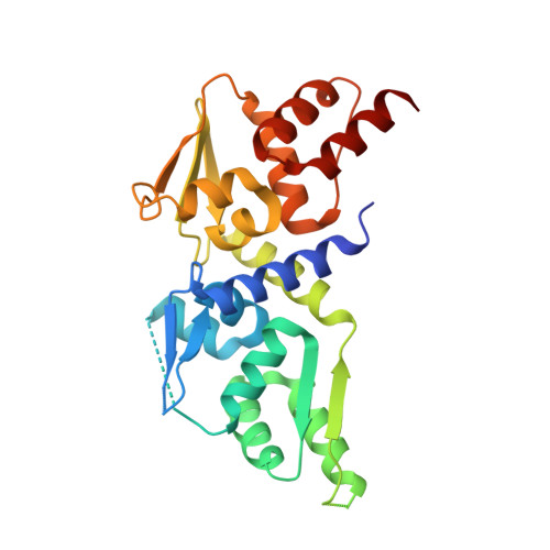
    The POZ domain is an evolutionarily conserved protein-protein interaction domain that is found in approximately 40 mammalian transcription factors. POZ domains mediate both homodimerization and the heteromeric interactions of different POZ-domain transcription factors with each other. Miz1 is a POZ-domain transcription factor that regulates cell-cycle arrest and DNA-damage responses. The activities of Miz1 are altered by its interaction with the POZ-domain transcriptional repressors BCL6 and NAC1, and these interactions have been implicated in tumourigenesis in B-cell lymphomas and in ovarian serous carcinomas that overexpress BCL6 and NAC1, respectively. A strategy for the purification of tethered POZ domains that form forced heterodimers is described, and crystal structures of the heterodimeric POZ domains of Miz1/BCL6 and of Miz1/NAC1 are reported. These structures will be relevant for the design of therapeutics that target POZ-domain interaction interfaces.


  • Organizational Affiliation
    • School of Biology, University of Leeds, Leeds LS2 9JT, England.

Macromolecule Content 

  • Total Structure Weight: 113.31 kDa 
  • Atom Count: 7,350 
  • Modeled Residue Count: 941 
  • Deposited Residue Count: 1,024 
  • Unique protein chains: 1

Macromolecules

Find similar proteins by:|  3D Structure
Entity ID: 1
MoleculeChains  Sequence LengthOrganismDetailsImage
Zinc finger and BTB domain-containing protein 17,B-cell lymphoma 6 protein
A, B, C, D
256Homo sapiensMutation(s): 0 
Gene Names: ZBTB17MIZ1ZNF151ZNF60BCL6BCL5LAZ3ZBTB27ZNF51
UniProt & NIH Common Fund Data Resources
Find proteins for P41182 (Homo sapiens)
Explore P41182 
Go to UniProtKB:  P41182
PHAROS:  P41182
GTEx:  ENSG00000113916 
Find proteins for Q13105 (Homo sapiens)
Explore Q13105 
Go to UniProtKB:  Q13105
PHAROS:  Q13105
GTEx:  ENSG00000116809 
Entity Groups
Sequence Clusters30% Identity50% Identity70% Identity90% Identity95% Identity100% Identity
UniProt GroupsQ13105P41182
Sequence Annotations
Expand
Reference Sequence

Experimental Data & Validation

Experimental Data

  • Method: X-RAY DIFFRACTION
  • Resolution: 2.23 Å
  • R-Value Free:  0.240 (Depositor), 0.234 (DCC) 
  • R-Value Work:  0.211 (Depositor) 
  • R-Value Observed: 0.213 (Depositor) 
Space Group: P 21 21 21
Unit Cell:
Length ( Å )Angle ( ˚ )
a = 91.85α = 90
b = 97.88β = 90
c = 119.88γ = 90
Software Package:
Software NamePurpose
Aimlessdata scaling
XDSdata reduction
PDB_EXTRACTdata extraction
PHASERphasing
ARPmodel building
REFMACrefinement
XSCALEdata reduction

Structure Validation

View Full Validation Report


Entry History 

& Funding Information

Deposition Data


Funding OrganizationLocationGrant Number
Yorkshire Cancer ResearchUnited KingdomL334

Revision History  (Full details and data files)

  • Version 1.0: 2014-12-10
    Type: Initial release
  • Version 1.1: 2014-12-17
    Changes: Database references
  • Version 1.2: 2024-05-08
    Changes: Data collection, Database references