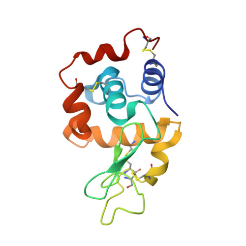
4RW1

Hen egg-white lysozyme structure from a spent-beam experiment at LCLS: original beam


Experimental Data Snapshot

  • Method: X-RAY DIFFRACTION
  • Resolution: 1.90 Å
  • R-Value Free: 0.256 
  • R-Value Work: 0.219 
  • R-Value Observed: 0.221 

wwPDB Validation   3D Report Full Report


This is version 1.3 of the entry. See complete history


Literature

Characterization and use of the spent beam for serial operation of LCLS.

Boutet, S.Foucar, L.Barends, T.R.Botha, S.Doak, R.B.Koglin, J.E.Messerschmidt, M.Nass, K.Schlichting, I.Seibert, M.M.Shoeman, R.L.Williams, G.J.

(2015) J Synchrotron Radiat 22: 634-643

  • DOI: https://doi.org/10.1107/S1600577515004002
  • Primary Citation of Related Structures:  
    4RW1, 4RW2

  • PubMed Abstract: 

    X-ray free-electron laser sources such as the Linac Coherent Light Source offer very exciting possibilities for unique research. However, beam time at such facilities is very limited and in high demand. This has led to significant efforts towards beam multiplexing of various forms. One such effort involves re-using the so-called spent beam that passes through the hole in an area detector after a weak interaction with a primary sample. This beam can be refocused into a secondary interaction region and used for a second, independent experiment operating in series. The beam profile of this refocused beam was characterized for a particular experimental geometry at the Coherent X-ray Imaging instrument at LCLS. A demonstration of this multiplexing capability was performed with two simultaneous serial femtosecond crystallography experiments, both yielding interpretable data of sufficient quality to produce electron density maps.


  • Organizational Affiliation

    Linac Coherent Light Source, SLAC National Accelerator Laboratory, 2575 Sand Hill Road, Menlo Park, CA 94025, USA.


Macromolecules
Find similar proteins by:  (by identity cutoff)  |  3D Structure
Entity ID: 1
MoleculeChains Sequence LengthOrganismDetailsImage
Lysozyme C129Gallus gallusMutation(s): 0 
EC: 3.2.1.17
UniProt
Find proteins for P00698 (Gallus gallus)
Explore P00698 
Go to UniProtKB:  P00698
Entity Groups  
Sequence Clusters30% Identity50% Identity70% Identity90% Identity95% Identity100% Identity
UniProt GroupP00698
Sequence Annotations
Expand
  • Reference Sequence
Experimental Data & Validation

Experimental Data

  • Method: X-RAY DIFFRACTION
  • Resolution: 1.90 Å
  • R-Value Free: 0.256 
  • R-Value Work: 0.219 
  • R-Value Observed: 0.221 
  • Space Group: P 43 21 2
Unit Cell:
Length ( Å )Angle ( ˚ )
a = 79.24α = 90
b = 79.24β = 90
c = 38.23γ = 90
Software Package:
Software NamePurpose
LCLSdata collection
REFMACrefinement
CrystFELdata reduction
CrystFELdata scaling
REFMACphasing

Structure Validation

View Full Validation Report


Entry History 

Deposition Data

Revision History  (Full details and data files)

  • Version 1.0: 2015-05-20
    Type: Initial release
  • Version 1.1: 2017-11-15
    Changes: Data collection
  • Version 1.2: 2018-02-14
    Changes: Data collection
  • Version 1.3: 2023-09-20
    Changes: Data collection, Database references, Derived calculations, Refinement description