4RV8 | pdb_00004rv8

Co-Crystal Structure of the Catalytic Domain of the Inosine Monophosphate Dehydrogenase from Cryptosporidium parvum and the inhibitor p131


Experimental Data Snapshot

  • Method: X-RAY DIFFRACTION
  • Resolution: 2.05 Å
  • R-Value Free: 
    0.229 (Depositor), 0.236 (DCC) 
  • R-Value Work: 
    0.187 (Depositor) 
  • R-Value Observed: 
    0.189 (Depositor) 

Starting Model: experimental
View more details

wwPDB Validation 3D Report Full Report

Validation slider image for 4RV8

Ligand Structure Quality Assessment 


This is version 1.5 of the entry. See complete history

Literature

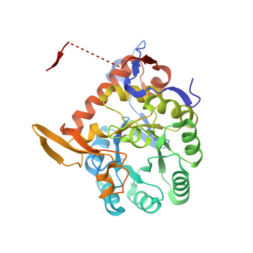
Structure of Cryptosporidium IMP dehydrogenase bound to an inhibitor with in vivo antiparasitic activity.

Kim, Y.Makowska-Grzyska, M.Gorla, S.K.Gollapalli, D.R.Cuny, G.D.Joachimiak, A.Hedstrom, L.

(2015) Acta Crystallogr F Struct Biol Commun 71: 531-538

  • DOI: https://doi.org/10.1107/S2053230X15000187
  • Primary Citation Related Structures: 
    4RV8

  • PubMed Abstract: 

    Inosine 5'-monophosphate dehydrogenase (IMPDH) is a promising target for the treatment of Cryptosporidium infections. Here, the structure of C. parvum IMPDH (CpIMPDH) in complex with inosine 5'-monophosphate (IMP) and P131, an inhibitor with in vivo anticryptosporidial activity, is reported. P131 contains two aromatic groups, one of which interacts with the hypoxanthine ring of IMP, while the second interacts with the aromatic ring of a tyrosine in the adjacent subunit. In addition, the amine and NO2 moieties bind in hydrated cavities, forming water-mediated hydrogen bonds to the protein. The design of compounds to replace these water molecules is a new strategy for the further optimization of C. parvum inhibitors for both antiparasitic and antibacterial applications.


  • Organizational Affiliation
    • Center for Structural Genomics of Infectious Diseases, Computational Institute, University of Chicago, 5735 S. Ellis Avenue, Chicago, IL 60637, USA.

Macromolecule Content 

  • Total Structure Weight: 157.93 kDa 
  • Atom Count: 10,414 
  • Modeled Residue Count: 1,307 
  • Deposited Residue Count: 1,444 
  • Unique protein chains: 1

Macromolecules

Find similar proteins by:|  3D Structure
Entity ID: 1
MoleculeChains  Sequence LengthOrganismDetailsImage
Inosine-5'-monophosphate dehydrogenase
A, B, C, D
361Cryptosporidium parvumMutation(s): 0 
Gene Names: 56k.02cgd6_20
EC: 1.1.1.205
UniProt
Find proteins for Q8T6T2 (Cryptosporidium parvum)
Explore Q8T6T2 
Go to UniProtKB:  Q8T6T2
Entity Groups
Sequence Clusters30% Identity50% Identity70% Identity90% Identity95% Identity100% Identity
UniProt GroupQ8T6T2
Sequence Annotations
Expand
Reference Sequence

Small Molecules

Ligands 4 Unique
IDChains Name / Formula / InChI Key2D Diagram3D Interactions
I13

Query on I13


Download:Ideal Coordinates CCD File
F [auth A],
J [auth B],
M [auth B],
P [auth C]
1-(2-{3-[(1E)-N-(2-aminoethoxy)ethanimidoyl]phenyl}propan-2-yl)-3-(4-chloro-3-nitrophenyl)urea
C20 H24 Cl N5 O4
MAIRRISEQUGPIE-DHRITJCHSA-N
IMP

Query on IMP


Download:Ideal Coordinates CCD File
E [auth A],
I [auth B],
N [auth C],
Q [auth D]
INOSINIC ACID
C10 H13 N4 O8 P
GRSZFWQUAKGDAV-KQYNXXCUSA-N
PEG

Query on PEG


Download:Ideal Coordinates CCD File
H [auth A],
L [auth B]
DI(HYDROXYETHYL)ETHER
C4 H10 O3
MTHSVFCYNBDYFN-UHFFFAOYSA-N
GOL

Query on GOL


Download:Ideal Coordinates CCD File
G [auth A],
K [auth B],
O [auth C],
R [auth D]
GLYCEROL
C3 H8 O3
PEDCQBHIVMGVHV-UHFFFAOYSA-N
Modified Residues  1 Unique
IDChains TypeFormula2D DiagramParent
OCS
Query on OCS
A, B, C, D
L-PEPTIDE LINKINGC3 H7 N O5 SCYS

Experimental Data & Validation

Experimental Data

  • Method: X-RAY DIFFRACTION
  • Resolution: 2.05 Å
  • R-Value Free:  0.229 (Depositor), 0.236 (DCC) 
  • R-Value Work:  0.187 (Depositor) 
  • R-Value Observed: 0.189 (Depositor) 
Space Group: P 1 21 1
Unit Cell:
Length ( Å )Angle ( ˚ )
a = 89.188α = 90
b = 91.832β = 103.75
c = 92.021γ = 90
Software Package:
Software NamePurpose
SBC-Collectdata collection
HKL-3000data collection
HKL-3000phasing
MOLREPphasing
PHENIXrefinement
HKL-3000data reduction
HKL-3000data scaling

Structure Validation

View Full Validation Report


Ligand Structure Quality Assessment 


Entry History 

Revision History  (Full details and data files)

  • Version 1.0: 2014-12-31
    Type: Initial release
  • Version 1.1: 2015-12-16
    Changes: Database references
  • Version 1.2: 2017-02-08
    Changes: Structure summary
  • Version 1.3: 2017-07-26
    Changes: Refinement description, Source and taxonomy
  • Version 1.4: 2023-09-20
    Changes: Data collection, Database references, Derived calculations, Refinement description
  • Version 1.5: 2023-12-06
    Changes: Data collection