Characterizing and Overriding the Structural Mechanism of the Quizartinib-Resistant FLT3 "Gatekeeper" F691L Mutation with PLX3397.
Smith, C.C., Zhang, C., Lin, K.C., Lasater, E.A., Zhang, Y., Massi, E., Damon, L.E., Pendleton, M., Bashir, A., Sebra, R., Perl, A., Kasarskis, A., Shellooe, R., Tsang, G., Carias, H., Powell, B., Burton, E.A., Matusow, B., Zhang, J., Spevak, W., Ibrahim, P.N., Le, M.H., Hsu, H.H., Habets, G., West, B.L., Bollag, G., Shah, N.P.(2015) Cancer Discov 5: 668-679
- PubMed: 25847190 Search on PubMedSearch on PubMed Central
- DOI: https://doi.org/10.1158/2159-8290.CD-15-0060
- Primary Citation Related Structures:
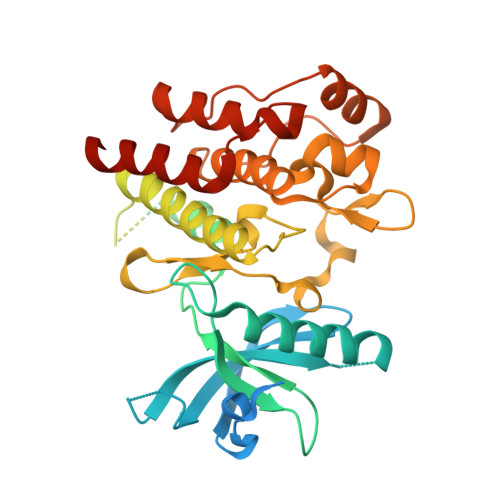
4RT7 - PubMed Abstract:
Tyrosine kinase domain mutations are a common cause of acquired clinical resistance to tyrosine kinase inhibitors (TKI) used to treat cancer, including the FLT3 inhibitor quizartinib. Mutation of kinase "gatekeeper" residues, which control access to an allosteric pocket adjacent to the ATP-binding site, has been frequently implicated in TKI resistance. The molecular underpinnings of gatekeeper mutation-mediated resistance are incompletely understood. We report the first cocrystal structure of FLT3 with the TKI quizartinib, which demonstrates that quizartinib binding relies on essential edge-to-face aromatic interactions with the gatekeeper F691 residue, and F830 within the highly conserved Asp-Phe-Gly motif in the activation loop. This reliance makes quizartinib critically vulnerable to gatekeeper and activation loop substitutions while minimizing the impact of mutations elsewhere. Moreover, we identify PLX3397, a novel FLT3 inhibitor that retains activity against the F691L mutant due to a binding mode that depends less vitally on specific interactions with the gatekeeper position. We report the first cocrystal structure of FLT3 with a kinase inhibitor, elucidating the structural mechanism of resistance due to the gatekeeper F691L mutation. PLX3397 is a novel FLT3 inhibitor with in vitro activity against this mutation but is vulnerable to kinase domain mutations in the FLT3 activation loop.
- Division of Hematology/Oncology, University of California, San Francisco, California. Helen Diller Family Comprehensive Cancer Center, University of California, San Francisco, California.
Organizational Affiliation: