4RLV | pdb_00004rlv

Crystal Structure of AnkB 24 Ankyrin Repeats in Complex with AnkR Autoinhibition Segment


Experimental Data Snapshot

  • Method: X-RAY DIFFRACTION
  • Resolution: 3.49 Å
  • R-Value Free: 
    0.253 (Depositor), 0.251 (DCC) 
  • R-Value Work: 
    0.220 (Depositor), 0.221 (DCC) 
  • R-Value Observed: 
    0.222 (Depositor) 

wwPDB Validation 3D Report Full Report

Validation slider image for 4RLV

This is version 1.4 of the entry. See complete history

Literature

Structural basis of diverse membrane target recognitions by ankyrins.

Wang, C.Wei, Z.Chen, K.Ye, F.Yu, C.Bennett, V.Zhang, M.

(2014) Elife 3: e04353

  • DOI: https://doi.org/10.7554/eLife.04353
  • Primary Citation Related Structures: 
    4RLV, 4RLY

  • PubMed Abstract: 

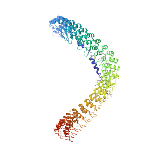
    Ankyrin adaptors together with their spectrin partners coordinate diverse ion channels and cell adhesion molecules within plasma membrane domains and thereby promote physiological activities including fast signaling in the heart and nervous system. Ankyrins specifically bind to numerous membrane targets through their 24 ankyrin repeats (ANK repeats), although the mechanism for the facile and independent evolution of these interactions has not been resolved. Here we report the structures of ANK repeats in complex with an inhibitory segment from the C-terminal regulatory domain and with a sodium channel Nav1.2 peptide, respectively, showing that the extended, extremely conserved inner groove spanning the entire ANK repeat solenoid contains multiple target binding sites capable of accommodating target proteins with very diverse sequences via combinatorial usage of these sites. These structures establish a framework for understanding the evolution of ankyrins' membrane targets, with implications for other proteins containing extended ANK repeat domains.


  • Organizational Affiliation
    • Division of Life Science, State Key Laboratory of Molecular Neuroscience, Hong Kong University of Science and Technology, Hong Kong, Hong Kong.

Macromolecule Content 

  • Total Structure Weight: 98.88 kDa 
  • Atom Count: 6,305 
  • Modeled Residue Count: 838 
  • Deposited Residue Count: 910 
  • Unique protein chains: 1

Macromolecules

Find similar proteins by:|  3D Structure
Entity ID: 1
MoleculeChains  Sequence LengthOrganismDetailsImage
Ankyrin-1, Ankyrin-2910Mus musculusHomo sapiens
This entity is chimeric
Mutation(s): 0 
Gene Names: Ank1ANK2
UniProt & NIH Common Fund Data Resources
Find proteins for Q01484 (Homo sapiens)
Explore Q01484 
Go to UniProtKB:  Q01484
PHAROS:  Q01484
GTEx:  ENSG00000145362 
Find proteins for Q02357 (Mus musculus)
Explore Q02357 
Go to UniProtKB:  Q02357
Entity Groups
Sequence Clusters30% Identity50% Identity70% Identity90% Identity95% Identity100% Identity
UniProt GroupsQ02357Q01484
Sequence Annotations
Expand
Reference Sequence

Small Molecules

Ligands 1 Unique
IDChains Name / Formula / InChI Key2D Diagram3D Interactions
SO4

Query on SO4


Download:Ideal Coordinates CCD File
B [auth A]
C [auth A]
D [auth A]
E [auth A]
F [auth A]
B [auth A],
C [auth A],
D [auth A],
E [auth A],
F [auth A],
G [auth A],
H [auth A],
I [auth A],
J [auth A]
SULFATE ION
O4 S
QAOWNCQODCNURD-UHFFFAOYSA-L
Modified Residues  1 Unique
IDChains TypeFormula2D DiagramParent
MSE
Query on MSE
A
L-PEPTIDE LINKINGC5 H11 N O2 SeMET

Experimental Data & Validation

Experimental Data

  • Method: X-RAY DIFFRACTION
  • Resolution: 3.49 Å
  • R-Value Free:  0.253 (Depositor), 0.251 (DCC) 
  • R-Value Work:  0.220 (Depositor), 0.221 (DCC) 
  • R-Value Observed: 0.222 (Depositor) 
Space Group: H 3 2
Unit Cell:
Length ( Å )Angle ( ˚ )
a = 179.685α = 90
b = 179.685β = 90
c = 304.856γ = 120
Software Package:
Software NamePurpose
DENZOdata reduction
SCALEPACKdata scaling
SHARPphasing
PHENIXrefinement
PDB_EXTRACTdata extraction
ADSCdata collection
HKL-2000data reduction
HKL-2000data scaling

Structure Validation

View Full Validation Report


Entry History 

Deposition Data

Revision History  (Full details and data files)

  • Version 1.0: 2014-11-26
    Type: Initial release
  • Version 1.1: 2015-05-20
    Changes: Database references
  • Version 1.2: 2017-08-02
    Changes: Source and taxonomy
  • Version 1.3: 2017-11-22
    Changes: Refinement description
  • Version 1.4: 2024-11-27
    Changes: Data collection, Database references, Derived calculations, Structure summary