4RJY | pdb_00004rjy

Crystal structure of E. coli L-Threonine Aldolase in complex with a non-covalently uncleaved bound L-serine substrate


Experimental Data Snapshot

  • Method: X-RAY DIFFRACTION
  • Resolution: 2.10 Å
  • R-Value Free: 
    0.279 (Depositor), 0.279 (DCC) 
  • R-Value Work: 
    0.212 (Depositor), 0.214 (DCC) 
  • R-Value Observed: 
    0.279 (Depositor) 

wwPDB Validation 3D Report Full Report

Validation slider image for 4RJY

This is version 1.3 of the entry. See complete history

Literature

Molecular basis of E. colil-threonine aldolase catalytic inactivation at low pH.

Remesh, S.G.Ghatge, M.S.Ahmed, M.H.Musayev, F.N.Gandhi, A.Chowdhury, N.di Salvo, M.L.Kellogg, G.E.Contestabile, R.Schirch, V.Safo, M.K.

(2015) Biochim Biophys Acta 1854: 278-283

  • DOI: https://doi.org/10.1016/j.bbapap.2014.12.023
  • Primary Citation Related Structures: 
    4RJY

  • PubMed Abstract: 

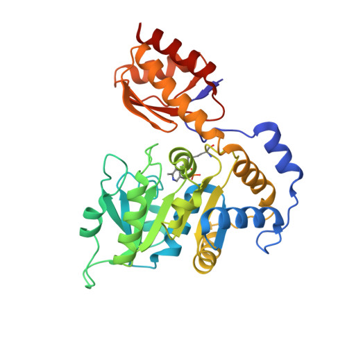
    L-Threonine aldolases (TAs), a family of enzymes belonging to the fold-type I pyridoxal 5'-phosphate (PLP) dependent enzymes, play a role in catalyzing the reversible cleavage of l-3-hydroxy-α-amino acids to glycine and the corresponding aldehydes. Threonine aldolases have great biotechnological potential for the syntheses of pharmaceutically relevant drug molecules because of their stereospecificity. The pH-dependency of their catalytic activity, affecting reaction intermediates, led us to study the effect of low-pH on Escherichia coli TA (eTA) structure. We report here a low-pH crystal structure of eTA at 2.1 Å resolution, with a non-covalently bound uncleaved l-serine substrate, and a PLP cofactor bound as an internal aldimine. This structure contrasts with other eTA structures obtained at physiological pH that show products or substrates bound as PLP-external aldimines. The non-productive binding at low-pH is due to an unusual substrate serine binding orientation in which the α-amino group and carboxylate group are in the wrong positions (relative to the active site residues) as a result of protonation of the α-amino group of the serine, as well as the active site histidines, His83 and His126. Protonation of these residues prevents the characteristic nucleophilic attack of the α-amino group of substrate serine on C4' of PLP to form the external aldimine. Our study shows that at low pH the change in charge distribution at the active site can result in substrates binding in a non-productive orientation.


  • Organizational Affiliation
    • Department of Medicinal Chemistry, School of Pharmacy, Institute for Structural Biology and Drug Discovery, Virginia Commonwealth University, Richmond, VA, USA.

Macromolecule Content 

  • Total Structure Weight: 147.59 kDa 
  • Atom Count: 11,165 
  • Modeled Residue Count: 1,328 
  • Deposited Residue Count: 1,332 
  • Unique protein chains: 1

Macromolecules

Find similar proteins by:|  3D Structure
Entity ID: 1
MoleculeChains  Sequence LengthOrganismDetailsImage
Low specificity L-threonine aldolase
A, B, C, D
333Escherichia coli KTE79Mutation(s): 0 
Gene Names: A1UU_02790

Small Molecules

Ligands 2 Unique
IDChains Name / Formula / InChI Key2D Diagram3D Interactions
SER

Query on SER


Download:Ideal Coordinates CCD File
F [auth A],
I [auth B],
K [auth C],
N [auth D]
SERINE
C3 H7 N O3
MTCFGRXMJLQNBG-REOHCLBHSA-N
NA

Query on NA


Download:Ideal Coordinates CCD File
E [auth A]
G [auth B]
H [auth B]
J [auth C]
L [auth D]
E [auth A],
G [auth B],
H [auth B],
J [auth C],
L [auth D],
M [auth D]
SODIUM ION
Na
FKNQFGJONOIPTF-UHFFFAOYSA-N
Modified Residues  1 Unique
IDChains TypeFormula2D DiagramParent
LLP
Query on LLP
A, B, C, D
L-PEPTIDE LINKINGC14 H22 N3 O7 PLYS

Experimental Data & Validation

Experimental Data

  • Method: X-RAY DIFFRACTION
  • Resolution: 2.10 Å
  • R-Value Free:  0.279 (Depositor), 0.279 (DCC) 
  • R-Value Work:  0.212 (Depositor), 0.214 (DCC) 
  • R-Value Observed: 0.279 (Depositor) 
Space Group: P 1 21 1
Unit Cell:
Length ( Å )Angle ( ˚ )
a = 77.16α = 90
b = 104.88β = 92.49
c = 84.91γ = 90
Software Package:
Software NamePurpose
d*TREKdata reduction
CNSrefinement
PDB_EXTRACTdata extraction
CrystalCleardata collection
d*TREKdata scaling
AMoREphasing

Structure Validation

View Full Validation Report


Entry History 

Deposition Data

Revision History  (Full details and data files)

  • Version 1.0: 2014-10-29
    Type: Initial release
  • Version 1.1: 2015-01-21
    Changes: Database references
  • Version 1.2: 2015-02-04
    Changes: Database references
  • Version 1.3: 2025-03-26
    Changes: Data collection, Database references, Derived calculations, Structure summary