4R4E

Structure of GlnR-DNA complex


Experimental Data Snapshot

  • Method: X-RAY DIFFRACTION
  • Resolution: 2.57 Å
  • R-Value Free: 0.275 
  • R-Value Work: 0.254 
  • R-Value Observed: 0.254 

wwPDB Validation   3D Report Full Report


Ligand Structure Quality Assessment 


This is version 1.2 of the entry. See complete history


Literature

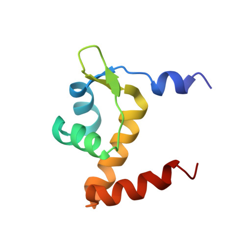
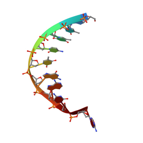
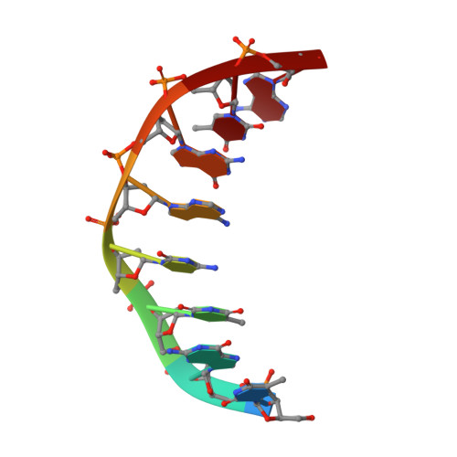
Structures of regulatory machinery reveal novel molecular mechanisms controlling B. subtilis nitrogen homeostasis.

Schumacher, M.A.Chinnam, N.B.Cuthbert, B.Tonthat, N.K.Whitfill, T.

(2015) Genes Dev 29: 451-464

  • DOI: https://doi.org/10.1101/gad.254714.114
  • Primary Citation of Related Structures:  
    4R22, 4R24, 4R25, 4R4E, 4RX6, 4S0R

  • PubMed Abstract: 

    All cells must sense and adapt to changing nutrient availability. However, detailed molecular mechanisms coordinating such regulatory pathways remain poorly understood. In Bacillus subtilis, nitrogen homeostasis is controlled by a unique circuitry composed of the regulator TnrA, which is deactivated by feedback-inhibited glutamine synthetase (GS) during nitrogen excess and stabilized by GlnK upon nitrogen depletion, and the repressor GlnR. Here we describe a complete molecular dissection of this network. TnrA and GlnR, the global nitrogen homeostatic transcription regulators, are revealed as founders of a new structural family of dimeric DNA-binding proteins with C-terminal, flexible, effector-binding sensors that modulate their dimerization. Remarkably, the TnrA sensor domains insert into GS intersubunit catalytic pores, destabilizing the TnrA dimer and causing an unprecedented GS dodecamer-to-tetradecamer conversion, which concomitantly deactivates GS. In contrast, each subunit of the GlnK trimer "templates" active TnrA dimers. Unlike TnrA, GlnR sensors mediate an autoinhibitory dimer-destabilizing interaction alleviated by GS, which acts as a GlnR chaperone. Thus, these studies unveil heretofore unseen mechanisms by which inducible sensor domains drive metabolic reprograming in the model Gram-positive bacterium B. subtilis.


  • Organizational Affiliation

    Department of Biochemistry, Duke University Medical Center, Durham, North Carolina 27710, USA maria.schumacher@duke.edu.


Macromolecules

Find similar proteins by:  (by identity cutoff)  |  3D Structure
Entity ID: 1
MoleculeChains Sequence LengthOrganismDetailsImage
HTH-type transcriptional regulator GlnRA [auth B],
B [auth A]
84Bacillus subtilis subsp. subtilis str. 168Mutation(s): 0 
Gene Names: glnRBSU17450
UniProt
Find proteins for P37582 (Bacillus subtilis (strain 168))
Explore P37582 
Go to UniProtKB:  P37582
Entity Groups  
Sequence Clusters30% Identity50% Identity70% Identity90% Identity95% Identity100% Identity
UniProt GroupP37582
Sequence Annotations
Expand
  • Reference Sequence

Find similar nucleic acids by:  Sequence   |   3D Structure  

Entity ID: 2
MoleculeChains LengthOrganismImage
DNA (5'-D(*AP*TP*TP*CP*TP*GP*AP*CP*A)-3')C [auth D]9synthetic construct
Sequence Annotations
Expand
  • Reference Sequence

Find similar nucleic acids by:  Sequence   |   3D Structure  

Entity ID: 3
MoleculeChains LengthOrganismImage
DNA (5'-D(*TP*GP*TP*CP*AP*GP*TP*A)-3')D [auth E]8synthetic construct
Sequence Annotations
Expand
  • Reference Sequence
Small Molecules
Ligands 1 Unique
IDChains Name / Formula / InChI Key2D Diagram3D Interactions
CXS
Query on CXS

Download Ideal Coordinates CCD File 
E [auth A]3-CYCLOHEXYL-1-PROPYLSULFONIC ACID
C9 H19 N O3 S
PJWWRFATQTVXHA-UHFFFAOYSA-N
Experimental Data & Validation

Experimental Data

  • Method: X-RAY DIFFRACTION
  • Resolution: 2.57 Å
  • R-Value Free: 0.275 
  • R-Value Work: 0.254 
  • R-Value Observed: 0.254 
  • Space Group: I 21 21 21
Unit Cell:
Length ( Å )Angle ( ˚ )
a = 76.35α = 90
b = 103.3β = 90
c = 137.14γ = 90
Software Package:
Software NamePurpose
CNSrefinement
PDB_EXTRACTdata extraction
ADSCdata collection
MOSFLMdata reduction
SCALAdata scaling
SOLVEphasing

Structure Validation

View Full Validation Report


Ligand Structure Quality Assessment 


Entry History 

Deposition Data

Revision History  (Full details and data files)

  • Version 1.0: 2015-03-04
    Type: Initial release
  • Version 1.1: 2024-02-28
    Changes: Data collection, Database references, Derived calculations
  • Version 1.2: 2024-04-03
    Changes: Refinement description