


Crystal Structure of an Anti-sigma Factor Antagonist from Mycobacterium paratuberculosis
Dranow, D.M., Clifton, M.C., Edwards, T.E., Lorimer, D.To be published.
Experimental Data Snapshot
wwPDB Validation 3D Report Full Report

Entity ID: 1 | |||||
|---|---|---|---|---|---|
| Molecule | Chains | Sequence Length | Organism | Details | Image |
| Anti-sigma factor antagonist | 144 | Mycobacterium avium subsp. paratuberculosis K-10 | Mutation(s): 0 Gene Names: MAP_0380 | 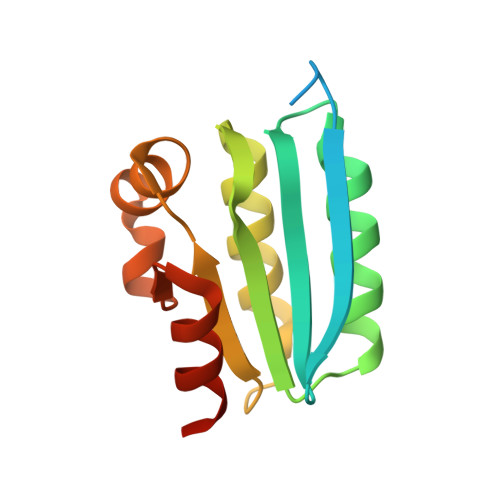 | |
UniProt | |||||
Entity Groups | |||||
| Sequence Clusters | 30% Identity50% Identity70% Identity90% Identity95% Identity100% Identity | ||||
| UniProt Group | Q744G1 | ||||
Sequence AnnotationsExpand | |||||
Reference Sequence | |||||
| Ligands 3 Unique | |||||
|---|---|---|---|---|---|
| ID | Chains | Name / Formula / InChI Key | 2D Diagram | 3D Interactions | |
| CIT Download:Ideal Coordinates CCD File | E [auth A], J [auth B], N [auth C], U [auth D] | CITRIC ACID C6 H8 O7 KRKNYBCHXYNGOX-UHFFFAOYSA-N |  | ||
| GOL Download:Ideal Coordinates CCD File | I [auth A], M [auth B], X [auth D] | GLYCEROL C3 H8 O3 PEDCQBHIVMGVHV-UHFFFAOYSA-N |  | ||
| EDO Download:Ideal Coordinates CCD File | F [auth A] G [auth A] H [auth A] K [auth B] L [auth B] | 1,2-ETHANEDIOL C2 H6 O2 LYCAIKOWRPUZTN-UHFFFAOYSA-N |  | ||
| Length ( Å ) | Angle ( ˚ ) |
|---|---|
| a = 58.17 | α = 69.66 |
| b = 58.3 | β = 78.61 |
| c = 58.9 | γ = 78.94 |
| Software Name | Purpose |
|---|---|
| XSCALE | data scaling |
| REFMAC | refinement |
| PDB_EXTRACT | data extraction |
| BALBES | phasing |


