Crystal structure of the mouse interleukin-3 beta-receptor: insights into interleukin-3 binding and receptor activation.
Carr, P.D., Ewens, C.L., Dai, J., Ollis, D.L., Murphy, J.M., Jackson, C.J., Young, I.G.(2014) Biochem J 463: 393-403
- PubMed: 25137390 Search on PubMed
- DOI: https://doi.org/10.1042/BJ20140863
- Primary Citation Related Structures:
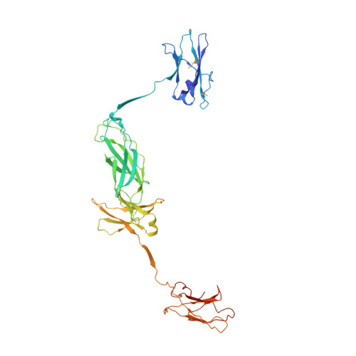
4QQV - PubMed Abstract:
Interleukin-3 (IL-3) is a cytokine secreted by mast cells and activated T-cells known to be an important regulator of differentiation, survival, proliferation and activation of a range of haemopoietic lineages. The effects of IL-3 on target cells are mediated by a transmembrane receptor system composed of a cytokine-specific α-subunit and a β-subunit, the principal signalling entity. In the mouse, two β-subunits have co-evolved: a common β-subunit (βc) shared between IL-3 and the related cytokines IL-5 and granulocyte/macrophage colony-stimulating factor (GM-CSF); and an IL-3-specific β-subunit (βIL-3). βIL-3 differs from βc in its specificity for IL-3 and its capacity to bind IL-3 directly in the absence of an α-subunit, and, in the absence of structural information, the basis for these properties has remained enigmatic. In the present study, we have solved the crystal structure of the βIL-3 ectodomain at 3.45 Å (1 Å=0.1 nm) resolution. This structure provides the first evidence that βIL-3 adopts an arch-shaped intertwined homodimer with similar topology to the paralogous βc structure. In contrast with apo-βc, however, the ligand-binding interface of βIL-3 appears to pre-exist in a conformation receptive to IL-3 engagement. Molecular modelling of the IL-3-βIL-3 interface, in conjunction with previous mutational studies, suggests that divergent evolution of both βIL-3 and IL-3 underlies their unique capacity for direct interaction and specificity.
- *Research School of Chemistry, Australian National University, Acton 0200, Australia.
Organizational Affiliation: