4QKH

Dimeric form of human LLT1, a ligand for NKR-P1


Experimental Data Snapshot

  • Method: X-RAY DIFFRACTION
  • Resolution: 1.80 Å
  • R-Value Free: 0.250 
  • R-Value Work: 0.175 
  • R-Value Observed: 0.180 

wwPDB Validation   3D Report Full Report


Ligand Structure Quality Assessment 


This is version 1.4 of the entry. See complete history


Literature

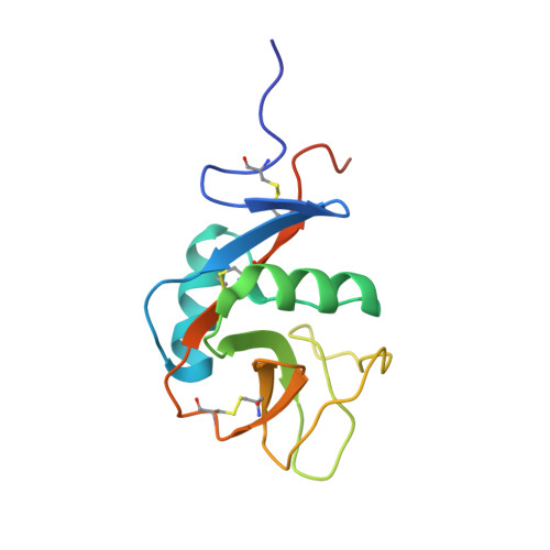
Four crystal structures of human LLT1, a ligand of human NKR-P1, in varied glycosylation and oligomerization states

Skalova, T.Blaha, J.Harlos, K.Duskova, J.Koval, T.Stransky, J.Hasek, J.Vanek, O.Dohnalek, J.

(2015) Acta Crystallogr D Biol Crystallogr 71: 578-591

  • DOI: https://doi.org/10.1107/S1399004714027928
  • Primary Citation of Related Structures:  
    4QKG, 4QKH, 4QKI, 4QKJ

  • PubMed Abstract: 
  • Human LLT1 is a C-type lectin-like ligand of NKR-P1 (CD161, gene KLRB1), a C-type lectin-like receptor of natural killer cells. Using X-ray diffraction, the first experimental structures of human LLT1 were determined. Four structures of LLT1 under various conditions were determined: monomeric, dimeric deglycosylated after the first N-acetylglucosamine unit in two forms and hexameric with homogeneous GlcNAc2Man5 glycosylation ...

    Human LLT1 is a C-type lectin-like ligand of NKR-P1 (CD161, gene KLRB1), a C-type lectin-like receptor of natural killer cells. Using X-ray diffraction, the first experimental structures of human LLT1 were determined. Four structures of LLT1 under various conditions were determined: monomeric, dimeric deglycosylated after the first N-acetylglucosamine unit in two forms and hexameric with homogeneous GlcNAc2Man5 glycosylation. The dimeric form follows the classical dimerization mode of human CD69. The monomeric form keeps the same fold with the exception of the position of an outer part of the long loop region. The hexamer of glycosylated LLT1 consists of three classical dimers. The hexameric packing may indicate a possible mode of interaction of C-type lectin-like proteins in the glycosylated form.


    Organizational Affiliation

    Institute of Biotechnology, Academy of Sciences of the Czech Republic, v.v.i., Vídeňská 1083, 142 20 Praha 4, Czech Republic.


Macromolecules
Find similar proteins by:  (by identity cutoff)  |  3D Structure
Entity ID: 1
MoleculeChainsSequence LengthOrganismDetailsImage
C-type lectin domain family 2 member DA, B135Homo sapiensMutation(s): 1 
Gene Names: CLAXCLEC2BCLEC2DLLT1OCIL
UniProt & NIH Common Fund Data Resources
Find proteins for Q9UHP7 (Homo sapiens)
Explore Q9UHP7 
Go to UniProtKB:  Q9UHP7
PHAROS:  Q9UHP7
Entity Groups  
Sequence Clusters30% Identity50% Identity70% Identity90% Identity95% Identity100% Identity
UniProt GroupQ9UHP7
Protein Feature View
Expand
  • Reference Sequence
Small Molecules
Ligands 1 Unique
IDChainsName / Formula / InChI Key2D Diagram3D Interactions
NAG
Query on NAG

Download Ideal Coordinates CCD File 
C [auth A],
D [auth A],
E [auth B],
F [auth B]
2-acetamido-2-deoxy-beta-D-glucopyranose
C8 H15 N O6
OVRNDRQMDRJTHS-FMDGEEDCSA-N
 Ligand Interaction
Experimental Data & Validation

Experimental Data

Unit Cell:
Length ( Å )Angle ( ˚ )
a = 50.87α = 90
b = 57.82β = 90
c = 82.25γ = 90
Software Package:
Software NamePurpose
BALBESphasing
REFMACrefinement
MOSFLMdata reduction
Aimlessdata scaling

Structure Validation

View Full Validation Report


Ligand Structure Quality Assessment 


Entry History 

Deposition Data

Revision History  (Full details and data files)

  • Version 1.0: 2015-03-11
    Type: Initial release
  • Version 1.1: 2016-01-27
    Changes: Database references
  • Version 1.2: 2018-06-13
    Changes: Data collection, Source and taxonomy
  • Version 1.3: 2018-06-20
    Changes: Data collection, Database references
  • Version 1.4: 2020-07-29
    Type: Remediation
    Reason: Carbohydrate remediation
    Changes: Data collection, Database references, Derived calculations, Structure summary