


Crystal structure of goat lactoperoxidase in complex with octopamine at 2.1 Angstrom resolution
Singh, R.P., Kushwaha, G.S., Singh, A.K., Sinha, M., Kaur, P., Sharma, S., Singh, T.P.To be published.
Experimental Data Snapshot
Entity ID: 1 | |||||
|---|---|---|---|---|---|
| Molecule | Chains | Sequence Length | Organism | Details | Image |
| Lactoperoxidase | 595 | Capra hircus | Mutation(s): 0 EC: 1.11.1.7 | 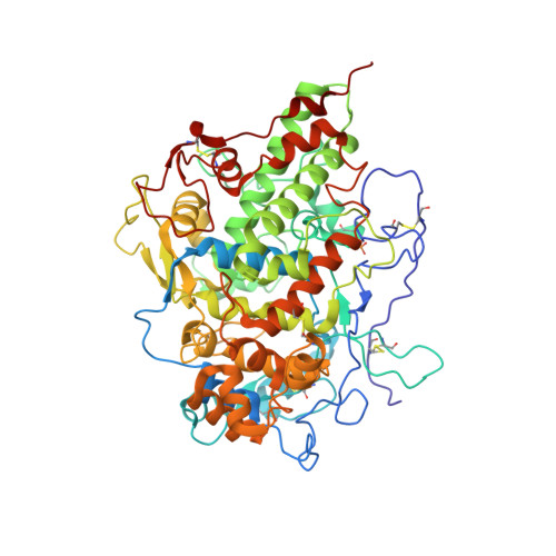 | |
UniProt | |||||
Entity Groups | |||||
| Sequence Clusters | 30% Identity50% Identity70% Identity90% Identity95% Identity100% Identity | ||||
| UniProt Group | A0A452E9Y6 | ||||
Glycosylation | |||||
| Glycosylation Sites: 4 | |||||
Sequence AnnotationsExpand | |||||
Reference Sequence | |||||
| Ligands 8 Unique | |||||
|---|---|---|---|---|---|
| ID | Chains | Name / Formula / InChI Key | 2D Diagram | 3D Interactions | |
| HEM Download:Ideal Coordinates CCD File | B [auth A] | PROTOPORPHYRIN IX CONTAINING FE C34 H32 Fe N4 O4 KABFMIBPWCXCRK-RGGAHWMASA-L |  | ||
| NAG Download:Ideal Coordinates CCD File | C [auth A], D [auth A], E [auth A], F [auth A] | 2-acetamido-2-deoxy-beta-D-glucopyranose C8 H15 N O6 OVRNDRQMDRJTHS-FMDGEEDCSA-N |  | ||
| OTR Download:Ideal Coordinates CCD File | O [auth A] | 4-(2R-AMINO-1-HYDROXYETHYL)PHENOL C8 H11 N O2 QHGUCRYDKWKLMG-QMMMGPOBSA-N |  | ||
| IOD Download:Ideal Coordinates CCD File | H [auth A] I [auth A] J [auth A] K [auth A] L [auth A] | IODIDE ION I XMBWDFGMSWQBCA-UHFFFAOYSA-M |  | ||
| PEG Download:Ideal Coordinates CCD File | P [auth A], Q [auth A] | DI(HYDROXYETHYL)ETHER C4 H10 O3 MTHSVFCYNBDYFN-UHFFFAOYSA-N |  | ||
| EDO Download:Ideal Coordinates CCD File | Y [auth A], Z [auth A] | 1,2-ETHANEDIOL C2 H6 O2 LYCAIKOWRPUZTN-UHFFFAOYSA-N |  | ||
| SCN Download:Ideal Coordinates CCD File | N [auth A] | THIOCYANATE ION C N S ZMZDMBWJUHKJPS-UHFFFAOYSA-M |  | ||
| CA Download:Ideal Coordinates CCD File | G [auth A] | CALCIUM ION Ca BHPQYMZQTOCNFJ-UHFFFAOYSA-N |  | ||
| Modified Residues 1 Unique | |||||
|---|---|---|---|---|---|
| ID | Chains | Type | Formula | 2D Diagram | Parent |
| SEP Query on SEP | A | L-PEPTIDE LINKING | C3 H8 N O6 P |  | SER |
| Length ( Å ) | Angle ( ˚ ) |
|---|---|
| a = 53.947 | α = 90 |
| b = 80.356 | β = 102.81 |
| c = 75.391 | γ = 90 |
| Software Name | Purpose |
|---|---|
| HKL-2000 | data collection |
| MOLREP | phasing |
| REFMAC | refinement |
| HKL-2000 | data reduction |
| HKL-2000 | data scaling |


