L-Arabinose Binding, Isomerization, and Epimerization by D-Xylose Isomerase: X-Ray/Neutron Crystallographic and Molecular Simulation Study.
Langan, P., Sangha, A.K., Wymore, T., Parks, J.M., Yang, Z.K., Hanson, B.L., Fisher, Z., Mason, S.A., Blakeley, M.P., Forsyth, V.T., Glusker, J.P., Carrell, H.L., Smith, J.C., Keen, D.A., Graham, D.E., Kovalevsky, A.(2014) Structure 22: 1287-1300
- PubMed: 25132082 Search on PubMed
- DOI: https://doi.org/10.1016/j.str.2014.07.002
- Primary Citation Related Structures:
4QDP, 4QDW, 4QE1, 4QE4, 4QE5, 4QEE, 4QEH - PubMed Abstract:
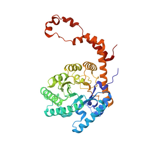
D-xylose isomerase (XI) is capable of sugar isomerization and slow conversion of some monosaccharides into their C2-epimers. We present X-ray and neutron crystallographic studies to locate H and D atoms during the respective isomerization and epimerization of L-arabinose to L-ribulose and L-ribose, respectively. Neutron structures in complex with cyclic and linear L-arabinose have demonstrated that the mechanism of ring-opening is the same as for the reaction with D-xylose. Structural evidence and QM/MM calculations show that in the reactive Michaelis complex L-arabinose is distorted to the high-energy (5)S1 conformation; this may explain the apparent high KM for this sugar. MD-FEP simulations indicate that amino acid substitutions in a hydrophobic pocket near C5 of L-arabinose can enhance sugar binding. L-ribulose and L-ribose were found in furanose forms when bound to XI. We propose that these complexes containing Ni(2+) cofactors are Michaelis-like and the isomerization between these two sugars proceeds via a cis-ene-diol mechanism.
- Biology and Soft Matter Division, Oak Ridge National Laboratory, Oak Ridge, TN 37831, USA.
Organizational Affiliation: