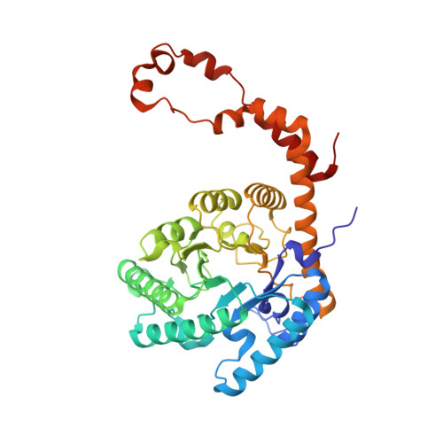
4QE1

Room temperature X-ray structure of D-xylose isomerase in complex with two Cd2+ ions and L-ribulose


Experimental Data Snapshot

  • Method: X-RAY DIFFRACTION
  • Resolution: 1.55 Å
  • R-Value Free: 0.190 
  • R-Value Work: 0.158 
  • R-Value Observed: 0.148 

wwPDB Validation   3D Report Full Report


Ligand Structure Quality Assessment 


This is version 1.3 of the entry. See complete history


Literature

L-Arabinose Binding, Isomerization, and Epimerization by D-Xylose Isomerase: X-Ray/Neutron Crystallographic and Molecular Simulation Study.

Langan, P.Sangha, A.K.Wymore, T.Parks, J.M.Yang, Z.K.Hanson, B.L.Fisher, Z.Mason, S.A.Blakeley, M.P.Forsyth, V.T.Glusker, J.P.Carrell, H.L.Smith, J.C.Keen, D.A.Graham, D.E.Kovalevsky, A.

(2014) Structure 22: 1287-1300

  • DOI: https://doi.org/10.1016/j.str.2014.07.002
  • Primary Citation of Related Structures:  
    4QDP, 4QDW, 4QE1, 4QE4, 4QE5, 4QEE, 4QEH

  • PubMed Abstract: 

    D-xylose isomerase (XI) is capable of sugar isomerization and slow conversion of some monosaccharides into their C2-epimers. We present X-ray and neutron crystallographic studies to locate H and D atoms during the respective isomerization and epimerization of L-arabinose to L-ribulose and L-ribose, respectively. Neutron structures in complex with cyclic and linear L-arabinose have demonstrated that the mechanism of ring-opening is the same as for the reaction with D-xylose. Structural evidence and QM/MM calculations show that in the reactive Michaelis complex L-arabinose is distorted to the high-energy (5)S1 conformation; this may explain the apparent high KM for this sugar. MD-FEP simulations indicate that amino acid substitutions in a hydrophobic pocket near C5 of L-arabinose can enhance sugar binding. L-ribulose and L-ribose were found in furanose forms when bound to XI. We propose that these complexes containing Ni(2+) cofactors are Michaelis-like and the isomerization between these two sugars proceeds via a cis-ene-diol mechanism.


  • Organizational Affiliation

    Biology and Soft Matter Division, Oak Ridge National Laboratory, Oak Ridge, TN 37831, USA.


Macromolecules
Find similar proteins by:  (by identity cutoff)  |  3D Structure
Entity ID: 1
MoleculeChains Sequence LengthOrganismDetailsImage
Xylose isomerase388Streptomyces rubiginosusMutation(s): 0 
EC: 5.3.1.5
UniProt
Find proteins for P24300 (Streptomyces rubiginosus)
Explore P24300 
Go to UniProtKB:  P24300
Entity Groups  
Sequence Clusters30% Identity50% Identity70% Identity90% Identity95% Identity100% Identity
UniProt GroupP24300
Sequence Annotations
Expand
  • Reference Sequence
Experimental Data & Validation

Experimental Data

  • Method: X-RAY DIFFRACTION
  • Resolution: 1.55 Å
  • R-Value Free: 0.190 
  • R-Value Work: 0.158 
  • R-Value Observed: 0.148 
  • Space Group: I 2 2 2
Unit Cell:
Length ( Å )Angle ( ˚ )
a = 93.865α = 90
b = 99.869β = 90
c = 102.84γ = 90
Software Package:
Software NamePurpose
HKL-3000data collection
PHASERphasing
SHELXL-97refinement
HKL-3000data reduction
HKL-3000data scaling

Structure Validation

View Full Validation Report


Ligand Structure Quality Assessment 


Entry History 

Deposition Data

Revision History  (Full details and data files)

  • Version 1.0: 2014-09-03
    Type: Initial release
  • Version 1.1: 2014-09-10
    Changes: Database references
  • Version 1.2: 2020-07-29
    Type: Remediation
    Reason: Carbohydrate remediation
    Changes: Data collection, Derived calculations
  • Version 1.3: 2024-02-28
    Changes: Data collection, Database references, Structure summary