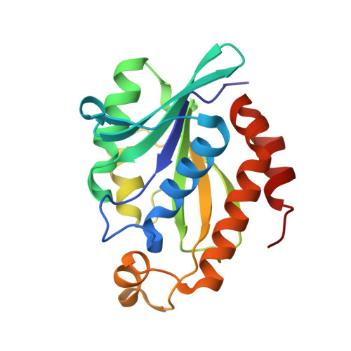
4QD3

Crystal structure of Peptidyl-tRNA hydrolase from Pseudomonas aeruginosa with 5-azacytidine at 1.89 Angstrom resolution


Experimental Data Snapshot

  • Method: X-RAY DIFFRACTION
  • Resolution: 1.89 Å
  • R-Value Free: 0.238 
  • R-Value Work: 0.186 
  • R-Value Observed: 0.189 

Starting Model: experimental
View more details

wwPDB Validation   3D Report Full Report


Ligand Structure Quality Assessment 


This is version 1.2 of the entry. See complete history


Literature

Structural and binding studies of peptidyl-tRNA hydrolase from Pseudomonas aeruginosa provide a platform for the structure-based inhibitor design against peptidyl-tRNA hydrolase

Singh, A.Kumar, A.Gautam, L.Sharma, P.Sinha, M.Bhushan, A.Kaur, P.Sharma, S.Arora, A.Singh, T.P.

(2014) Biochem J 463: 329-337

  • DOI: https://doi.org/10.1042/BJ20140631
  • Primary Citation of Related Structures:  
    4FNO, 4JC4, 4QAJ, 4QBK, 4QD3

  • PubMed Abstract: 

    During the course of protein synthesis in the cell, the translation process is often terminated due to various reasons. As a result, peptidyl-tRNA molecules are released which are toxic to the cell as well reducing the availability of free amino acid and tRNA molecules for the required protein synthesis in the cell. Such a situation is corrected by an enzyme, Pth (peptidyl-tRNA hydrolase), which catalyses the release of free tRNA and peptide moieties from peptidyl-tRNAs. This means that the active Pth is essential for the survival of bacteria. In order to design inhibitors of PaPth (Pth from Pseudomonas aeruginosa), we determined the structures of PaPth in its native and bound states with compounds amino acylate-tRNA analogue and 5-azacytidine. The structure determination of the native protein revealed that the substrate-binding site was partially occupied by Glu161 from the neigh-bouring molecule. The structure of PaPth indicated that the substrate-binding site can be broadly divided into three distinct subsites. The structures of the two complexes showed that the amino acylate-tRNA analogue filled three subsites, whereas 5-azacytidine filled two subsites. The common sugar and the base moieties of the two compounds occupied identical positions in the cleft. Using surface plasmon resonance, the dissociation constants for the amino acylate-tRNA analogue and 5-azacytidine were found to be 3.53×10⁻⁸ M and 5.82×10⁻⁸ M respectively.


  • Organizational Affiliation

    *Department of Biophysics, All India Institute of Medical Sciences, Ansari Nagar, New Delhi, 110029 Delhi, India.


Macromolecules
Find similar proteins by:  (by identity cutoff)  |  3D Structure
Entity ID: 1
MoleculeChains Sequence LengthOrganismDetailsImage
Peptidyl-tRNA hydrolase194Pseudomonas aeruginosa PAO1Mutation(s): 0 
Gene Names: PA4672pth
EC: 3.1.1.29
UniProt
Find proteins for Q9HVC3 (Pseudomonas aeruginosa (strain ATCC 15692 / DSM 22644 / CIP 104116 / JCM 14847 / LMG 12228 / 1C / PRS 101 / PAO1))
Explore Q9HVC3 
Go to UniProtKB:  Q9HVC3
Entity Groups  
Sequence Clusters30% Identity50% Identity70% Identity90% Identity95% Identity100% Identity
UniProt GroupQ9HVC3
Sequence Annotations
Expand
  • Reference Sequence
Small Molecules
Ligands 2 Unique
IDChains Name / Formula / InChI Key2D Diagram3D Interactions
5AE
Query on 5AE

Download Ideal Coordinates CCD File 
B [auth A]4-amino-1-(beta-D-ribofuranosyl)-1,3,5-triazin-2(1H)-one
C8 H12 N4 O5
NMUSYJAQQFHJEW-KVTDHHQDSA-N
GOL
Query on GOL

Download Ideal Coordinates CCD File 
C [auth A]GLYCEROL
C3 H8 O3
PEDCQBHIVMGVHV-UHFFFAOYSA-N
Experimental Data & Validation

Experimental Data

  • Method: X-RAY DIFFRACTION
  • Resolution: 1.89 Å
  • R-Value Free: 0.238 
  • R-Value Work: 0.186 
  • R-Value Observed: 0.189 
  • Space Group: P 61 2 2
Unit Cell:
Length ( Å )Angle ( ˚ )
a = 64.111α = 90
b = 64.111β = 90
c = 156.62γ = 120
Software Package:
Software NamePurpose
HKL-2000data collection
AMoREphasing
REFMACrefinement
HKL-2000data reduction
SCALEPACKdata scaling

Structure Validation

View Full Validation Report


Ligand Structure Quality Assessment 


Entry History 

Deposition Data

Revision History  (Full details and data files)

  • Version 1.0: 2014-06-25
    Type: Initial release
  • Version 1.1: 2014-11-12
    Changes: Database references
  • Version 1.2: 2023-11-08
    Changes: Data collection, Database references, Derived calculations, Refinement description