4QB9 | pdb_00004qb9

Crystal structure of Mycobacterium smegmatis Eis in complex with paromomycin


Experimental Data Snapshot

  • Method: X-RAY DIFFRACTION
  • Resolution: 3.29 Å
  • R-Value Free: 
    0.234 (Depositor), 0.230 (DCC) 
  • R-Value Work: 
    0.151 (Depositor), 0.163 (DCC) 
  • R-Value Observed: 
    0.156 (Depositor) 

wwPDB Validation 3D Report Full Report

Validation slider image for 4QB9

Ligand Structure Quality Assessment 


This is version 1.2 of the entry. See complete history

Literature

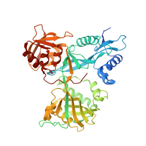
Structure of Mycobacterium smegmatis Eis in complex with paromomycin.

Kim, K.H.An, D.R.Yoon, H.J.Yang, J.K.Suh, S.W.

(2014) Acta Crystallogr Sect F Struct Biol Cryst Commun 70: 1173-1179

  • DOI: https://doi.org/10.1107/S2053230X14017385
  • Primary Citation Related Structures: 
    4QB9

  • PubMed Abstract: 

    The Rv2416c gene of Mycobacterium tuberculosis (Mtb) encodes the enhanced intracellular survival (Eis) protein that enhances intracellular survival of the pathogen in host macrophages during infection. The Mtb Eis protein is released into the cytoplasm of the phagocyte during intracellular infection and modulates the host immune response. It also contributes to drug resistance by acetylating multiple amine groups of aminoglycosides. Interestingly, the nonpathogenic M. smegmatis (Msm) contains a homologous eis gene (MSMEG_3513). The overall structures of Mtb Eis and Msm Eis are highly similar to each other, reflecting the high level (58%) of amino-acid sequence identity between them. Both Mtb Eis and Msm Eis are active as aminoglycoside acetyltransferases, while only Mtb Eis functions as an N(ℇ)-acetyltransferase to acetylate Lys55 of dual-specificity protein phosphatase 16 (DUSP16)/mitogen-activated protein kinase phosphatase 7 (MKP-7), leading to the suppression of host immune responses. Here, the crystal structure of Msm Eis in the paromomycin-bound form is reported, revealing detailed interactions between an aminoglycoside antibiotic and Msm Eis. The crystal structure of Msm Eis in the paromomycin-bound form has been determined at 3.3 Å resolution. This work provides potentially useful information for structure-guided discovery of Eis inhibitors as a novel antituberculosis drug against drug-resistant Mtb.


  • Organizational Affiliation
    • Department of Chemistry, College of Natural Sciences, Seoul National University, Seoul 151-742, Republic of Korea.

Macromolecule Content 

  • Total Structure Weight: 281.07 kDa 
  • Atom Count: 18,910 
  • Modeled Residue Count: 2,412 
  • Deposited Residue Count: 2,532 
  • Unique protein chains: 1

Macromolecules

Find similar proteins by:|  3D Structure
Entity ID: 1
MoleculeChains  Sequence LengthOrganismDetailsImage
Enhanced intracellular survival protein
A, B, C, D, E
A, B, C, D, E, F
422Mycolicibacterium smegmatis MC2 155Mutation(s): 0 
Gene Names: eisMSMEG_3513MSMEI_3433
UniProt
Find proteins for A0QY29 (Mycolicibacterium smegmatis (strain ATCC 700084 / mc(2)155))
Explore A0QY29 
Go to UniProtKB:  A0QY29
Entity Groups
Sequence Clusters30% Identity50% Identity70% Identity90% Identity95% Identity100% Identity
UniProt GroupA0QY29
Sequence Annotations
Expand
Reference Sequence

Small Molecules

Ligands 2 Unique
IDChains Name / Formula / InChI Key2D Diagram3D Interactions
PAR

Query on PAR


Download:Ideal Coordinates CCD File
G [auth A]
I [auth B]
K [auth C]
M [auth D]
O [auth E]
G [auth A],
I [auth B],
K [auth C],
M [auth D],
O [auth E],
Q [auth F]
PAROMOMYCIN
C23 H45 N5 O14
UOZODPSAJZTQNH-LSWIJEOBSA-N
SO4

Query on SO4


Download:Ideal Coordinates CCD File
H [auth A]
J [auth B]
L [auth C]
N [auth D]
P [auth E]
H [auth A],
J [auth B],
L [auth C],
N [auth D],
P [auth E],
R [auth F]
SULFATE ION
O4 S
QAOWNCQODCNURD-UHFFFAOYSA-L

Experimental Data & Validation

Experimental Data

  • Method: X-RAY DIFFRACTION
  • Resolution: 3.29 Å
  • R-Value Free:  0.234 (Depositor), 0.230 (DCC) 
  • R-Value Work:  0.151 (Depositor), 0.163 (DCC) 
  • R-Value Observed: 0.156 (Depositor) 
Space Group: P 21 21 21
Unit Cell:
Length ( Å )Angle ( ˚ )
a = 107.269α = 90
b = 126.536β = 90
c = 236.641γ = 90
Software Package:
Software NamePurpose
PHENIXrefinement
PDB_EXTRACTdata extraction

Structure Validation

View Full Validation Report


Ligand Structure Quality Assessment 


Entry History 

Deposition Data

Revision History  (Full details and data files)

  • Version 1.0: 2015-04-01
    Type: Initial release
  • Version 1.1: 2016-09-28
    Changes: Structure summary
  • Version 1.2: 2024-03-20
    Changes: Data collection, Database references, Derived calculations, Structure summary