4Q9U

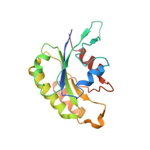
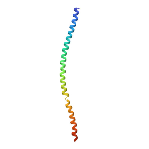
Crystal structure of the Rab5, Rabex-5delta and Rabaptin-5C21 complex


Experimental Data Snapshot

  • Method: X-RAY DIFFRACTION
  • Resolution: 4.62 Å
  • R-Value Free: 0.343 
  • R-Value Work: 0.251 
  • R-Value Observed: 0.256 

wwPDB Validation   3D Report Full Report


This is version 1.2 of the entry. See complete history


Literature

Molecular mechanism for Rabex-5 GEF activation by Rabaptin-5

Zhang, Z.Zhang, T.Wang, S.Gong, Z.Tang, C.Chen, J.Ding, J.

(2014) Elife 3: e02687-e02687

  • DOI: 10.7554/eLife.02687
  • Primary Citation of Related Structures:  
    4N3X, 4N3Y, 4N3Z, 4Q9U

  • PubMed Abstract: 
  • Rabex-5 and Rabaptin-5 function together to activate Rab5 and further promote early endosomal fusion in endocytosis. The Rabex-5 GEF activity is autoinhibited by the Rabex-5 CC domain (Rabex-5CC) and activated by the Rabaptin-5 C2-1 domain (Rabaptin-5C21) with yet unknown mechanism ...

    Rabex-5 and Rabaptin-5 function together to activate Rab5 and further promote early endosomal fusion in endocytosis. The Rabex-5 GEF activity is autoinhibited by the Rabex-5 CC domain (Rabex-5CC) and activated by the Rabaptin-5 C2-1 domain (Rabaptin-5C21) with yet unknown mechanism. We report here the crystal structures of Rabex-5 in complex with the dimeric Rabaptin-5C21 (Rabaptin-5C212) and in complex with Rabaptin-5C212 and Rab5, along with biophysical and biochemical analyses. We show that Rabex-5CC assumes an amphipathic α-helix which binds weakly to the substrate-binding site of the GEF domain, leading to weak autoinhibition of the GEF activity. Binding of Rabaptin-5C21 to Rabex-5 displaces Rabex-5CC to yield a largely exposed substrate-binding site, leading to release of the GEF activity. In the ternary complex the substrate-binding site of Rabex-5 is completely exposed to bind and activate Rab5. Our results reveal the molecular mechanism for the regulation of the Rabex-5 GEF activity.


    Organizational Affiliation

    State Key Laboratory of Molecular Biology, Institute of Biochemistry and Cell Biology, Shanghai Institutes for Biological Sciences, Chinese Academy of Sciences, Shanghai, China.


Macromolecules
Find similar proteins by:  (by identity cutoff)  |  3D Structure
Entity ID: 1
MoleculeChainsSequence LengthOrganismDetailsImage
Rab5 GDP/GTP exchange factorA, E317Homo sapiensMutation(s): 0 
Gene Names: RABGEF1RABEX5
UniProt
Find proteins for Q9UJ41 (Homo sapiens)
Explore Q9UJ41 
Go to UniProtKB:  Q9UJ41
Entity Groups  
Sequence Clusters30% Identity50% Identity70% Identity90% Identity95% Identity100% Identity
UniProt GroupQ9UJ41
Protein Feature View
Expand
  • Reference Sequence
Find similar proteins by:  (by identity cutoff)  |  3D Structure
Entity ID: 2
MoleculeChainsSequence LengthOrganismDetailsImage
Ras-related protein Rab-5AB, F171Homo sapiensMutation(s): 0 
Gene Names: RAB5ARAB5
EC: 3.6.5.2
UniProt & NIH Common Fund Data Resources
Find proteins for P20339 (Homo sapiens)
Explore P20339 
Go to UniProtKB:  P20339
PHAROS:  P20339
Entity Groups  
Sequence Clusters30% Identity50% Identity70% Identity90% Identity95% Identity100% Identity
UniProt GroupP20339
Protein Feature View
Expand
  • Reference Sequence
Find similar proteins by:  (by identity cutoff)  |  3D Structure
Entity ID: 3
MoleculeChainsSequence LengthOrganismDetailsImage
Rab GTPase-binding effector protein 1C, D, G, H92Homo sapiensMutation(s): 0 
Gene Names: RABEP1RAB5EPRABPT5RABPT5A
UniProt & NIH Common Fund Data Resources
Find proteins for Q15276 (Homo sapiens)
Explore Q15276 
Go to UniProtKB:  Q15276
PHAROS:  Q15276
Entity Groups  
Sequence Clusters30% Identity50% Identity70% Identity90% Identity95% Identity100% Identity
UniProt GroupQ15276
Protein Feature View
Expand
  • Reference Sequence
Experimental Data & Validation

Experimental Data

  • Method: X-RAY DIFFRACTION
  • Resolution: 4.62 Å
  • R-Value Free: 0.343 
  • R-Value Work: 0.251 
  • R-Value Observed: 0.256 
  • Space Group: P 41 21 2
Unit Cell:
Length ( Å )Angle ( ˚ )
a = 174.821α = 90
b = 174.821β = 90
c = 148.995γ = 90
Software Package:
Software NamePurpose
DENZOdata reduction
SCALEPACKdata scaling
PHASERphasing
PHENIXrefinement
PDB_EXTRACTdata extraction
HKL-2000data collection

Structure Validation

View Full Validation Report


Entry History 

Deposition Data

Revision History  (Full details and data files)

  • Version 1.0: 2014-07-23
    Type: Initial release
  • Version 1.1: 2015-12-09
    Changes: Database references
  • Version 1.2: 2017-11-22
    Changes: Refinement description