4Q31 | pdb_00004q31

The crystal structure of cystathione gamma lyase (CalE6) from Micromonospora echinospora


Experimental Data Snapshot

  • Method: X-RAY DIFFRACTION
  • Resolution: 2.10 Å
  • R-Value Free: 
    0.191 (Depositor), 0.190 (DCC) 
  • R-Value Work: 
    0.152 (Depositor), 0.152 (DCC) 
  • R-Value Observed: 
    0.153 (Depositor) 

wwPDB Validation 3D Report Full Report

Validation slider image for 4Q31

This is version 1.2 of the entry. See complete history

Literature

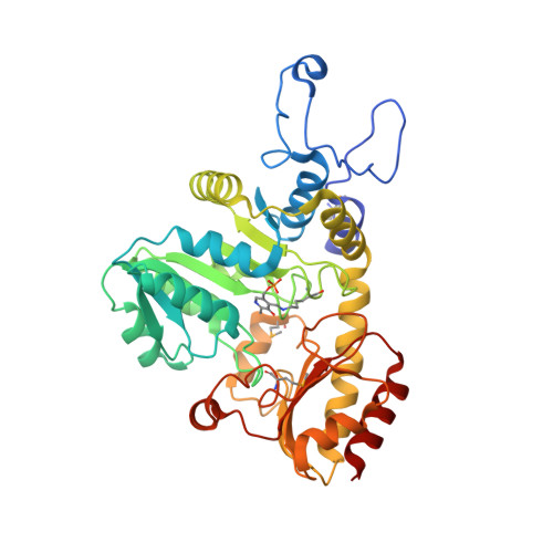
Structural dynamics of a methionine gamma-lyase for calicheamicin biosynthesis: Rotation of the conserved tyrosine stacking with pyridoxal phosphate.

Cao, H.Tan, K.Wang, F.Bigelow, L.Yennamalli, R.M.Jedrzejczak, R.Babnigg, G.Bingman, C.A.Joachimiak, A.Kharel, M.K.Singh, S.Thorson, J.S.Phillips, G.N.

(2016) Struct Dyn 3: 034702-034702

  • DOI: https://doi.org/10.1063/1.4948539
  • Primary Citation Related Structures: 
    4Q31

  • PubMed Abstract: 

    CalE6 from Micromonospora echinospora is a (pyridoxal 5' phosphate) PLP-dependent methionine γ-lyase involved in the biosynthesis of calicheamicins. We report the crystal structure of a CalE6 2-(N-morpholino)ethanesulfonic acid complex showing ligand-induced rotation of Tyr100, which stacks with PLP, resembling the corresponding tyrosine rotation of true catalytic intermediates of CalE6 homologs. Elastic network modeling and crystallographic ensemble refinement reveal mobility of the N-terminal loop, which involves both tetrameric assembly and PLP binding. Modeling and comparative structural analysis of PLP-dependent enzymes involved in Cys/Met metabolism shine light on the functional implications of the intrinsic dynamic properties of CalE6 in catalysis and holoenzyme maturation.


  • Organizational Affiliation
    • Biosciences at Rice, Rice University , 6100 Main St., Houston, Texas 77005, USA.

Macromolecule Content 

  • Total Structure Weight: 335.29 kDa 
  • Atom Count: 24,719 
  • Modeled Residue Count: 3,018 
  • Deposited Residue Count: 3,072 
  • Unique protein chains: 1

Macromolecules

Find similar proteins by:|  3D Structure
Entity ID: 1
MoleculeChains  Sequence LengthOrganismDetailsImage
cystathione gamma lyase CalE6
A, B, C, D, E
A, B, C, D, E, F, G, H
384Micromonospora echinosporaMutation(s): 1 
Gene Names: calE6
EC: 4.4.1.2
UniProt
Find proteins for Q8KNG3 (Micromonospora echinospora)
Explore Q8KNG3 
Go to UniProtKB:  Q8KNG3
Entity Groups
Sequence Clusters30% Identity50% Identity70% Identity90% Identity95% Identity100% Identity
UniProt GroupQ8KNG3
Sequence Annotations
Expand
Reference Sequence

Small Molecules

Ligands 4 Unique
IDChains Name / Formula / InChI Key2D Diagram3D Interactions
MES

Query on MES


Download:Ideal Coordinates CCD File
DA [auth D]
EB [auth H]
I [auth A]
KA [auth E]
Q [auth B]
DA [auth D],
EB [auth H],
I [auth A],
KA [auth E],
Q [auth B],
QA [auth F],
WA [auth G],
Y [auth C]
2-(N-MORPHOLINO)-ETHANESULFONIC ACID
C6 H13 N O4 S
SXGZJKUKBWWHRA-UHFFFAOYSA-N
GOL

Query on GOL


Download:Ideal Coordinates CCD File
AB [auth G]
BA [auth C]
BB [auth G]
CB [auth G]
GA [auth D]
AB [auth G],
BA [auth C],
BB [auth G],
CB [auth G],
GA [auth D],
HA [auth D],
HB [auth H],
IA [auth D],
JA [auth D],
L [auth A],
M [auth A],
MA [auth E],
N [auth A],
NA [auth E],
O [auth A],
OA [auth E],
PA [auth E],
S [auth B],
SA [auth F],
T [auth B],
TA [auth F],
U [auth B],
UA [auth F],
V [auth B],
W [auth B],
X [auth B],
YA [auth G],
ZA [auth G]
GLYCEROL
C3 H8 O3
PEDCQBHIVMGVHV-UHFFFAOYSA-N
FMT

Query on FMT


Download:Ideal Coordinates CCD File
CA [auth C],
DB [auth G],
IB [auth H],
P [auth A],
VA [auth F]
FORMIC ACID
C H2 O2
BDAGIHXWWSANSR-UHFFFAOYSA-N
CL

Query on CL


Download:Ideal Coordinates CCD File
AA [auth C]
EA [auth D]
FA [auth D]
FB [auth H]
GB [auth H]
AA [auth C],
EA [auth D],
FA [auth D],
FB [auth H],
GB [auth H],
J [auth A],
K [auth A],
LA [auth E],
R [auth B],
RA [auth F],
XA [auth G],
Z [auth C]
CHLORIDE ION
Cl
VEXZGXHMUGYJMC-UHFFFAOYSA-M
Modified Residues  2 Unique
IDChains TypeFormula2D DiagramParent
LLP
Query on LLP
A, B, C, D, E
A, B, C, D, E, F, G, H
L-PEPTIDE LINKINGC14 H22 N3 O7 PLYS
MSE
Query on MSE
A, B, C, D, E
A, B, C, D, E, F, G, H
L-PEPTIDE LINKINGC5 H11 N O2 SeMET

Experimental Data & Validation

Experimental Data

  • Method: X-RAY DIFFRACTION
  • Resolution: 2.10 Å
  • R-Value Free:  0.191 (Depositor), 0.190 (DCC) 
  • R-Value Work:  0.152 (Depositor), 0.152 (DCC) 
  • R-Value Observed: 0.153 (Depositor) 
Space Group: I 2 2 2
Unit Cell:
Length ( Å )Angle ( ˚ )
a = 146.855α = 90
b = 146.981β = 90
c = 349.905γ = 90
Software Package:
Software NamePurpose
SBC-Collectdata collection
SHELXDphasing
MLPHAREphasing
DMmodel building
ARPmodel building
WARPmodel building
HKL-3000phasing
PHENIXrefinement
HKL-3000data reduction
HKL-3000data scaling
DMphasing

Structure Validation

View Full Validation Report


Entry History 

Revision History  (Full details and data files)

  • Version 1.0: 2014-05-07
    Type: Initial release
  • Version 1.1: 2017-03-08
    Changes: Database references
  • Version 1.2: 2025-03-26
    Changes: Data collection, Database references, Derived calculations, Structure summary