4PRU

Crystal structure of dimethyllysine hen egg-white lysozyme in complex with sclx4 at 2.2 A resolution


Experimental Data Snapshot

  • Method: X-RAY DIFFRACTION
  • Resolution: 2.20 Å
  • R-Value Free: 0.235 
  • R-Value Work: 0.180 
  • R-Value Observed: 0.183 

wwPDB Validation   3D Report Full Report


Ligand Structure Quality Assessment 


This is version 1.2 of the entry. See complete history


Literature

Structural study of a small molecule receptor bound to dimethyllysine in lysozyme.

McGovern, R.E.Snarr, B.D.Lyons, J.A.McFarlane, J.Whiting, A.L.Paci, I.Hof, F.Crowley, P.B.

(2015) Chem Sci 6: 442-449

  • DOI: https://doi.org/10.1039/C4SC02383H
  • Primary Citation of Related Structures:  
    4N0J, 4PRU

  • PubMed Abstract: 

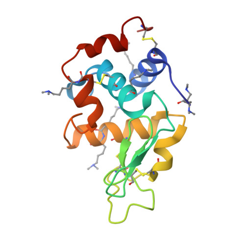
    Lysine is a ubiquitous residue on protein surfaces. Post translational modifications of lysine, including methylation to the mono-, di- or trimethylated amine result in chemical and structural alterations that have major consequences for protein interactions and signalling pathways. Small molecules that bind to methylated lysines are potential tools to modify such pathways. To make progress in this direction, detailed structural data of ligands in complex with methylated lysine is required. Here, we report a crystal structure of p -sulfonatocalix[4]arene (sclx 4 ) bound to methylated lysozyme in which the lysine residues were chemically modified from Lys-NH 3 + to Lys-NH(Me 2 ) + . Of the six possible dimethyllysine sites, sclx 4 selected Lys116-Me 2 and the dimethylamino substituent was deeply buried in the calixarene cavity. This complex confirms the tendency for Lys-Me 2 residues to form cation-π interactions, which have been shown to be important in protein recognition of histone tails bearing methylated lysines. Supporting data from NMR spectroscopy and MD simulations confirm the selectivity for Lys116-Me 2 in solution. The structure presented here may serve as a stepping stone to the development of new biochemical reagents that target methylated lysines.


  • Organizational Affiliation

    School of Chemistry, National University of Ireland Galway, University Road, Galway, Ireland.


Macromolecules
Find similar proteins by:  (by identity cutoff)  |  3D Structure
Entity ID: 1
MoleculeChains Sequence LengthOrganismDetailsImage
Lysozyme C
A, B
129Gallus gallusMutation(s): 0 
EC: 3.2.1.17
UniProt
Find proteins for P00698 (Gallus gallus)
Explore P00698 
Go to UniProtKB:  P00698
Entity Groups  
Sequence Clusters30% Identity50% Identity70% Identity90% Identity95% Identity100% Identity
UniProt GroupP00698
Sequence Annotations
Expand
  • Reference Sequence
Small Molecules
Modified Residues  2 Unique
IDChains TypeFormula2D DiagramParent
DM0
Query on DM0
A, B
L-PEPTIDE LINKINGC10 H22 N2 O2LYS
MLY
Query on MLY
A, B
L-PEPTIDE LINKINGC8 H18 N2 O2LYS
Experimental Data & Validation

Experimental Data

  • Method: X-RAY DIFFRACTION
  • Resolution: 2.20 Å
  • R-Value Free: 0.235 
  • R-Value Work: 0.180 
  • R-Value Observed: 0.183 
  • Space Group: P 1 21 1
Unit Cell:
Length ( Å )Angle ( ˚ )
a = 46.04α = 90
b = 30.28β = 96.62
c = 95.01γ = 90
Software Package:
Software NamePurpose
PHASERphasing
REFMACrefinement
XDSdata reduction
XSCALEdata scaling

Structure Validation

View Full Validation Report


Ligand Structure Quality Assessment 


Entry History 

Deposition Data

Revision History  (Full details and data files)

  • Version 1.0: 2014-11-12
    Type: Initial release
  • Version 1.1: 2015-01-21
    Changes: Database references
  • Version 1.2: 2023-09-20
    Changes: Data collection, Database references, Derived calculations, Refinement description