4P9R

Speciation of a group I intron into a lariat capping ribozyme (Heavy atom derivative)


Experimental Data Snapshot

  • Method: X-RAY DIFFRACTION
  • Resolution: 2.70 Å
  • R-Value Free: 0.256 
  • R-Value Work: 0.210 
  • R-Value Observed: 0.212 

wwPDB Validation   3D Report Full Report


This is version 1.5 of the entry. See complete history


Literature

Speciation of a group I intron into a lariat capping ribozyme.

Meyer, M.Nielsen, H.Olieric, V.Roblin, P.Johansen, S.D.Westhof, E.Masquida, B.

(2014) Proc Natl Acad Sci U S A 111: 7659-7664

  • DOI: https://doi.org/10.1073/pnas.1322248111
  • Primary Citation of Related Structures:  
    4P8Z, 4P9R, 6GYV

  • PubMed Abstract: 

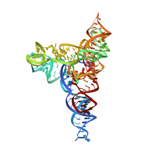
    The lariat-capping (LC) ribozyme is a natural ribozyme isolated from eukaryotic microorganisms. Despite apparent structural similarity to group I introns, the LC ribozyme catalyzes cleavage by a 2',5' branching reaction, leaving the 3' product with a 3-nt lariat cap that functionally substitutes for a conventional mRNA cap in the downstream pre-mRNA encoding a homing endonuclease. We describe the crystal structures of the precleavage and postcleavage LC ribozymes, which suggest that structural features inherited from group I ribozymes have undergone speciation due to profound changes in molecular selection pressure, ultimately giving rise to an original branching ribozyme family. The structures elucidate the role of key elements that regulate the activity of the LC ribozyme by conformational switching and suggest a mechanism by which the signal for branching is transmitted to the catalytic core. The structures also show how conserved interactions twist residues, forming the lariat to join chemical groups involved in branching.


  • Organizational Affiliation

    Architecture et Réactivité de l'ARN, Unité Propre de Recherche 9002, Institut de Biologie Moléculaire et Cellulaire, Université de Strasbourg, Centre National de la Recherche Scientifique, 67084 Strasbourg, France;


Macromolecules
Find similar nucleic acids by:  (by identity cutoff)  |  3D Structure
Entity ID: 1
MoleculeChains LengthOrganismImage
RNA (189-MER)192Didymium iridis
Sequence Annotations
Expand
  • Reference Sequence
Small Molecules
Ligands 2 Unique
IDChains Name / Formula / InChI Key2D Diagram3D Interactions
IRI
Query on IRI

Download Ideal Coordinates CCD File 
B [auth A]
C [auth A]
D [auth A]
E [auth A]
F [auth A]
B [auth A],
C [auth A],
D [auth A],
E [auth A],
F [auth A],
G [auth A],
H [auth A],
I [auth A],
J [auth A],
K [auth A]
IRIDIUM HEXAMMINE ION
H18 Ir N6
CGMAOQLDNKCXGK-UHFFFAOYSA-N
MG
Query on MG

Download Ideal Coordinates CCD File 
L [auth A],
M [auth A],
N [auth A]
MAGNESIUM ION
Mg
JLVVSXFLKOJNIY-UHFFFAOYSA-N
Experimental Data & Validation

Experimental Data

  • Method: X-RAY DIFFRACTION
  • Resolution: 2.70 Å
  • R-Value Free: 0.256 
  • R-Value Work: 0.210 
  • R-Value Observed: 0.212 
  • Space Group: P 21 21 21
Unit Cell:
Length ( Å )Angle ( ˚ )
a = 59.44α = 90
b = 86.26β = 90
c = 109.78γ = 90
Software Package:
Software NamePurpose
PHENIXrefinement

Structure Validation

View Full Validation Report


Entry History & Funding Information

Deposition Data


Funding OrganizationLocationGrant Number
French National Research AgencyFranceANR-10-BLAN-1502-02

Revision History  (Full details and data files)

  • Version 1.0: 2014-05-28
    Type: Initial release
  • Version 1.1: 2014-10-01
    Changes: Database references
  • Version 1.2: 2015-01-07
    Changes: Database references
  • Version 1.3: 2015-02-04
    Changes: Derived calculations
  • Version 1.4: 2017-10-04
    Changes: Author supporting evidence, Database references, Derived calculations, Other, Source and taxonomy
  • Version 1.5: 2023-12-27
    Changes: Data collection, Database references, Derived calculations, Refinement description