Structural Insights into Higher Order Assembly and Function of the Bacterial Microcompartment Protein PduA.
Pang, A., Frank, S., Brown, I., Warren, M.J., Pickersgill, R.W.(2014) J Biological Chem 289: 22377-22384
- PubMed: 24873823 Search on PubMedSearch on PubMed Central
- DOI: https://doi.org/10.1074/jbc.M114.569285
- Primary Citation Related Structures:
4P7T, 4P7V - PubMed Abstract:
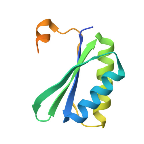
Bacterial microcompartments are large proteinaceous assemblies that are found in the cytoplasm of some bacteria. These structures consist of proteins constituting a shell that houses a number of enzymes involved in specific metabolic processes. The 1,2-propanediol-utilizing microcompartment is assembled from seven different types of shell proteins, one of which is PduA. It is one of the more abundant components of the shell and intriguingly can form nanotubule-like structures when expressed on its own in the cytoplasm of Escherichia coli. We propose a model that accounts for the size and appearance of these PduA structures and underpin our model using a combinatorial approach. Making strategic mutations at Lys-26, Val-51, and Arg-79, we targeted residues predicted to be important for PduA assembly. We present the effect of the amino acid residue substitution on the phenotype of the PduA higher order assemblies (transmission electron microscopy) and the crystal structure of the K26D mutant with one glycerol molecule bound to the central pore. Our results support the view that the hexamer-hexamer interactions seen in PduA crystals persist in the cytoplasmic structures and reveal the profound influence of the two key amino acids, Lys-26 and Arg-79, on tiling, not only in the crystal lattice but also in the bacterial cytoplasm. Understanding and controlling PduA assemblies is valuable in order to inform manipulation for synthetic biology and biotechnological applications.
- From the School of Biological and Chemical Sciences, Queen Mary University of London, Mile End Road, London E1 4NS, United Kingdom and.
Organizational Affiliation: