4OWJ

Crystal Structure of the Vibrio vulnificus Hemolysin/Cytolysin Beta-Trefoil Lectin


Experimental Data Snapshot

  • Method: X-RAY DIFFRACTION
  • Resolution: 2.00 Å
  • R-Value Free: 0.200 
  • R-Value Work: 0.166 
  • R-Value Observed: 0.168 

wwPDB Validation   3D Report Full Report


This is version 1.7 of the entry. See complete history


Literature

Glycan Specificity of the Vibrio vulnificus Hemolysin Lectin Outlines Evolutionary History of Membrane Targeting by a Toxin Family.

Kaus, K.Lary, J.W.Cole, J.L.Olson, R.

(2014) J Mol Biol 426: 2800-2812

  • DOI: https://doi.org/10.1016/j.jmb.2014.05.021
  • Primary Citation of Related Structures:  
    4OWJ, 4OWL

  • PubMed Abstract: 

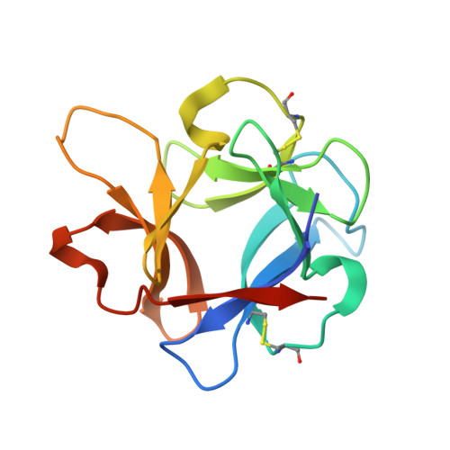
    Pore-forming toxins (PFTs) are a class of pathogen-secreted molecules that oligomerize to form transmembrane channels in cellular membranes. Determining the mechanism for how PFTs bind membranes is important in understanding their role in disease and for developing possible ways to block their action. Vibrio vulnificus, an aquatic pathogen responsible for severe food poisoning and septicemia in humans, secretes a PFT called V. vulnificus hemolysin (VVH), which contains a single C-terminal targeting domain predicted to resemble a β-trefoil lectin fold. In order to understand the selectivity of the lectin for glycan motifs, we expressed the isolated VVH β-trefoil domain and used glycan-chip screening to identify that VVH displays a preference for terminal galactosyl groups including N-acetyl-d-galactosamine and N-acetyl-d-lactosamine. The X-ray crystal structure of the VVH lectin domain solved to 2.0Å resolution reveals a heptameric ring arrangement similar to the oligomeric form of the related, but inactive, lectin from Vibrio cholerae cytolysin. Structures bound to glycerol, N-acetyl-d-galactosamine, and N-acetyl-d-lactosamine outline a common and versatile mode of recognition allowing VVH to target a wide variety of cell-surface ligands. Sequence analysis in light of our structural and functional data suggests that VVH may represent an earlier step in the evolution of Vibrio PFTs.


  • Organizational Affiliation

    Department of Molecular Biology and Biochemistry, Wesleyan University, 52 Lawn Avenue, Middletown, CT 06459, USA.


Macromolecules
Find similar proteins by:  (by identity cutoff)  |  3D Structure
Entity ID: 1
MoleculeChains Sequence LengthOrganismDetailsImage
Cytolysin
A, B, C, D, E
A, B, C, D, E, F, G
138Vibrio vulnificus CMCP6Mutation(s): 0 
Gene Names: VV2_0404vvhA
UniProt
Find proteins for P19247 (Vibrio vulnificus (strain CMCP6))
Explore P19247 
Go to UniProtKB:  P19247
Entity Groups  
Sequence Clusters30% Identity50% Identity70% Identity90% Identity95% Identity100% Identity
UniProt GroupP19247
Sequence Annotations
Expand
  • Reference Sequence
Small Molecules
Ligands 1 Unique
IDChains Name / Formula / InChI Key2D Diagram3D Interactions
GOL
Query on GOL

Download Ideal Coordinates CCD File 
H [auth A]
I [auth A]
J [auth B]
K [auth B]
L [auth C]
H [auth A],
I [auth A],
J [auth B],
K [auth B],
L [auth C],
M [auth C],
N [auth D],
O [auth D],
P [auth E],
Q [auth E],
R [auth F],
S [auth F],
T [auth G],
U [auth G]
GLYCEROL
C3 H8 O3
PEDCQBHIVMGVHV-UHFFFAOYSA-N
Experimental Data & Validation

Experimental Data

  • Method: X-RAY DIFFRACTION
  • Resolution: 2.00 Å
  • R-Value Free: 0.200 
  • R-Value Work: 0.166 
  • R-Value Observed: 0.168 
  • Space Group: P 21 21 21
Unit Cell:
Length ( Å )Angle ( ˚ )
a = 75.833α = 90
b = 118.214β = 90
c = 149.302γ = 90
Software Package:
Software NamePurpose
HKL-2000data reduction
PHASERphasing
PHENIXrefinement
PDB_EXTRACTdata extraction
HKL-2000data scaling
SCALEPACKdata scaling

Structure Validation

View Full Validation Report


Entry History & Funding Information

Deposition Data


Funding OrganizationLocationGrant Number
National Institutes of Health/National Institute Of Allergy and Infectious Diseases (NIH/NIAID)United StatesR15AI101977

Revision History  (Full details and data files)

  • Version 1.0: 2014-05-28
    Type: Initial release
  • Version 1.1: 2014-07-30
    Changes: Database references
  • Version 1.2: 2015-02-25
    Changes: Derived calculations
  • Version 1.3: 2015-10-07
    Changes: Other
  • Version 1.4: 2017-09-27
    Changes: Author supporting evidence, Derived calculations
  • Version 1.5: 2017-11-01
    Changes: Derived calculations
  • Version 1.6: 2019-12-11
    Changes: Author supporting evidence
  • Version 1.7: 2023-12-27
    Changes: Data collection, Database references