Structural Analysis of Replication Protein A Recruitment of the DNA Damage Response Protein SMARCAL1.
Feldkamp, M.D., Mason, A.C., Eichman, B.F., Chazin, W.J.(2014) Biochemistry 53: 3052-3061
- PubMed: 24730652 Search on PubMedSearch on PubMed Central
- DOI: https://doi.org/10.1021/bi500252w
- Primary Citation Related Structures:
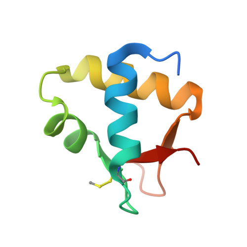
4OU0 - PubMed Abstract:
SWI/SNF-related, matrix-associated, actin-dependent regulator of chromatin, subfamily A-like1 (SMARCAL1) is a recently identified DNA damage response protein involved in remodeling stalled replication forks. The eukaryotic single-strand DNA binding protein replication protein A (RPA) recruits SMARCAL1 to stalled forks in vivo and facilitates regression of forks containing leading strand gaps. Both activities are mediated by a direct interaction between an RPA binding motif (RBM) at the N-terminus of SMARCAL1 and the C-terminal winged-helix domain of the RPA 32 kDa subunit (RPA32C). Here we report a biophysical and structural characterization of the SMARCAL1-RPA interaction. Isothermal titration calorimetry and circular dichroism spectroscopy revealed that RPA32C binds SMARCAL1-RBM with a Kd of 2.5 μM and induces a disorder-to-helix transition. The crystal structure of RPA32C was refined to 1.4 Å resolution, and the SMARCAL1-RBM binding site was mapped on the structure on the basis of nuclear magnetic resonance chemical shift perturbations. Conservation of the interaction surface to other RBM-containing proteins allowed construction of a model for the RPA32C/SMARCAL1-RBM complex. The implications of our results are discussed with respect to the recruitment of SMARCAL1 and other DNA damage response and repair proteins to stalled replication forks.
- Department of Biochemistry, ‡Department of Biological Sciences, §Department of Chemistry, and ∥Center for Structural Biology, Vanderbilt University , Nashville, Tennessee 37232, United States.
Organizational Affiliation: