4O6X | pdb_00004o6x

Crystal structure of human Ankyrin G death domain


Experimental Data Snapshot

  • Method: X-RAY DIFFRACTION
  • Resolution: 2.10 Å
  • R-Value Free: 
    0.231 (Depositor), 0.236 (DCC) 
  • R-Value Work: 
    0.182 (Depositor), 0.183 (DCC) 
  • R-Value Observed: 
    0.185 (Depositor) 

Starting Model: experimental
View more details

wwPDB Validation 3D Report Full Report

Validation slider image for 4O6X

This is version 1.3 of the entry. See complete history

Literature

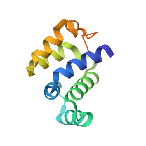
Crystal structure of human Ankyrin G death domain.

Liu, Y.Zhang, Y.Wang, J.H.

(2014) Proteins 82: 3476-3482

  • DOI: https://doi.org/10.1002/prot.24702
  • Primary Citation Related Structures: 
    4O6X

  • PubMed Abstract: 

    Ankyrins (Ank) are a ubiquitously expressed family of multifunctional membrane adapter proteins. Ankyrin G (AnkG) is critical for assembling and maintenance of the axon initial segment. Here we present the 2.1 Å crystal structure of human AnkG death domain (hAnkG-DD). The core death domain is composed of six α-helices and three 3₁₀-helices. It forms a hydrophobic pocket on the surface of the molecule. The C-terminal tail of the hAnkG-DD curves back to have the aromatic ring of a phenylalanine residue, Phe100 insert into this pocket, which anchors the flexible tail onto the core domain. Related DDs were selected for structure comparison. The major variations are at the C-terminal region, including the α6 and the long C-terminal extension. The results of size exclusion chromatography and analytical ultracentrifugation suggest that hAnkG-DD exists as monomer in solution. Our work should help for the future investigation of the structure-function of AnkG.


  • Organizational Affiliation
    • State Key Laboratory of Biomembrane and Membrane Biotechnology, College of Life Sciences, Peking University, Beijing, 100871, China.

Macromolecule Content 

  • Total Structure Weight: 27.64 kDa 
  • Atom Count: 1,606 
  • Modeled Residue Count: 193 
  • Deposited Residue Count: 242 
  • Unique protein chains: 1

Macromolecules

Find similar proteins by:|  3D Structure
Entity ID: 1
MoleculeChains  Sequence LengthOrganismDetailsImage
Ankyrin-3
A, B
121Homo sapiensMutation(s): 0 
Gene Names: ANK3Ankyrin G
UniProt & NIH Common Fund Data Resources
Find proteins for Q12955 (Homo sapiens)
Explore Q12955 
Go to UniProtKB:  Q12955
PHAROS:  Q12955
GTEx:  ENSG00000151150 
Entity Groups
Sequence Clusters30% Identity50% Identity70% Identity90% Identity95% Identity100% Identity
UniProt GroupQ12955
Sequence Annotations
Expand
Reference Sequence

Experimental Data & Validation

Experimental Data

  • Method: X-RAY DIFFRACTION
  • Resolution: 2.10 Å
  • R-Value Free:  0.231 (Depositor), 0.236 (DCC) 
  • R-Value Work:  0.182 (Depositor), 0.183 (DCC) 
  • R-Value Observed: 0.185 (Depositor) 
Space Group: P 21 21 21
Unit Cell:
Length ( Å )Angle ( ˚ )
a = 44.452α = 90
b = 56.817β = 90
c = 81.949γ = 90
Software Package:
Software NamePurpose
DENZOdata reduction
SCALEPACKdata scaling
MOLREPphasing
PHENIXrefinement
PDB_EXTRACTdata extraction
ADSCdata collection

Structure Validation

View Full Validation Report


Entry History 

Deposition Data

Revision History  (Full details and data files)

  • Version 1.0: 2014-10-29
    Type: Initial release
  • Version 1.1: 2014-12-17
    Changes: Database references
  • Version 1.2: 2015-02-11
    Changes: Other
  • Version 1.3: 2023-09-20
    Changes: Data collection, Database references, Refinement description