4O6F

Structural Basis of Estrogen Receptor Alpha Methylation Mediated by Histone Methyltransferase SmyD2


Experimental Data Snapshot

  • Method: X-RAY DIFFRACTION
  • Resolution: 2.82 Å
  • R-Value Free: 0.235 
  • R-Value Work: 0.175 
  • R-Value Observed: 0.178 

wwPDB Validation   3D Report Full Report


Ligand Structure Quality Assessment 


This is version 1.2 of the entry. See complete history


Literature

Structural Insights into Estrogen Receptor alpha Methylation by Histone Methyltransferase SMYD2, a Cellular Event Implicated in Estrogen Signaling Regulation.

Jiang, Y.Trescott, L.Holcomb, J.Zhang, X.Brunzelle, J.Sirinupong, N.Shi, X.Yang, Z.

(2014) J Mol Biol 426: 3413-3425

  • DOI: https://doi.org/10.1016/j.jmb.2014.02.019
  • Primary Citation of Related Structures:  
    4O6F

  • PubMed Abstract: 

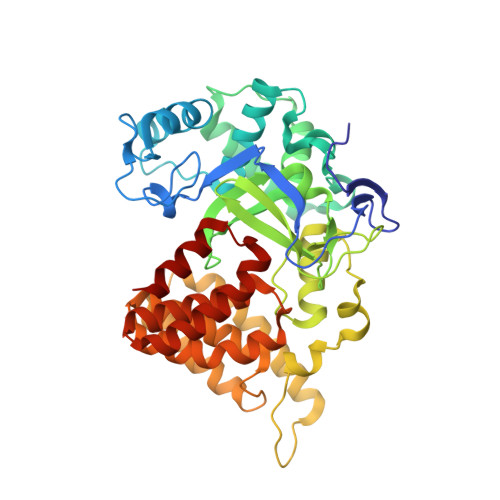
    Estrogen receptor (ER) signaling plays a pivotal role in many developmental processes and has been implicated in numerous diseases including cancers. We recently showed that direct ERα methylation by the multi-specificity histone lysine methyltransferase SMYD2 regulates estrogen signaling through repressing ERα-dependent transactivation. However, the mechanism controlling the specificity of the SMYD2-ERα interaction and the structural basis of SMYD2 substrate binding diversity are unknown. Here we present the crystal structure of SMYD2 in complex with a target lysine (Lys266)-containing ERα peptide. The structure reveals that ERα binds SMYD2 in a U-shaped conformation with the binding specificity determined mainly by residues C-terminal to the target lysine. The structure also reveals numerous intrapeptide contacts that ensure shape complementarity between the substrate and the active site of the enzyme, thereby likely serving as an additional structural determinant of substrate specificity. In addition, comparison of the SMYD2-ERα and SMYD2-p53 structures provides the first structural insight into the diverse nature of SMYD2 substrate recognition and suggests that the broad specificity of SMYD2 is achieved by multiple molecular mechanisms such as distinct peptide binding modes and the intrinsic dynamics of peptide ligands. Strikingly, a novel potentially SMYD2-specific polyethylene glycol binding site is identified in the CTD domain, implicating possible functions in extended substrate binding or protein-protein interactions. Our study thus provides the structural basis for the SMYD2-mediated ERα methylation, and the resulting knowledge of SMYD2 substrate specificity and target binding diversity could have important implications in selective drug design against a wide range of ERα-related diseases.


  • Organizational Affiliation

    Department of Biochemistry and Molecular Biology, Wayne State University School of Medicine, Detroit, MI 48201, USA.


Macromolecules
Find similar proteins by:  (by identity cutoff)  |  3D Structure
Entity ID: 1
MoleculeChains Sequence LengthOrganismDetailsImage
N-lysine methyltransferase SMYD2433Homo sapiensMutation(s): 1 
Gene Names: KMT3CSMYD2
EC: 2.1.1 (PDB Primary Data), 2.1.1.43 (PDB Primary Data)
UniProt & NIH Common Fund Data Resources
Find proteins for Q9NRG4 (Homo sapiens)
Explore Q9NRG4 
Go to UniProtKB:  Q9NRG4
PHAROS:  Q9NRG4
GTEx:  ENSG00000143499 
Entity Groups  
Sequence Clusters30% Identity50% Identity70% Identity90% Identity95% Identity100% Identity
UniProt GroupQ9NRG4
Sequence Annotations
Expand
  • Reference Sequence

Find similar proteins by:  Sequence   |   3D Structure  

Entity ID: 2
MoleculeChains Sequence LengthOrganismDetailsImage
Estrogen receptor11Homo sapiensMutation(s): 0 
UniProt & NIH Common Fund Data Resources
Find proteins for P03372 (Homo sapiens)
Explore P03372 
Go to UniProtKB:  P03372
PHAROS:  P03372
GTEx:  ENSG00000091831 
Entity Groups  
Sequence Clusters30% Identity50% Identity70% Identity90% Identity95% Identity100% Identity
UniProt GroupP03372
Sequence Annotations
Expand
  • Reference Sequence
Small Molecules
Ligands 4 Unique
IDChains Name / Formula / InChI Key2D Diagram3D Interactions
SAH
Query on SAH

Download Ideal Coordinates CCD File 
F [auth A]S-ADENOSYL-L-HOMOCYSTEINE
C14 H20 N6 O5 S
ZJUKTBDSGOFHSH-WFMPWKQPSA-N
PE8
Query on PE8

Download Ideal Coordinates CCD File 
G [auth A]3,6,9,12,15,18,21-HEPTAOXATRICOSANE-1,23-DIOL
C16 H34 O9
GLZWNFNQMJAZGY-UHFFFAOYSA-N
ZN
Query on ZN

Download Ideal Coordinates CCD File 
C [auth A],
D [auth A],
E [auth A]
ZINC ION
Zn
PTFCDOFLOPIGGS-UHFFFAOYSA-N
NI
Query on NI

Download Ideal Coordinates CCD File 
H [auth A],
I [auth A]
NICKEL (II) ION
Ni
VEQPNABPJHWNSG-UHFFFAOYSA-N
Binding Affinity Annotations 
IDSourceBinding Affinity
SAH BindingDB:  4O6F IC50: 180 (nM) from 1 assay(s)
Experimental Data & Validation

Experimental Data

  • Method: X-RAY DIFFRACTION
  • Resolution: 2.82 Å
  • R-Value Free: 0.235 
  • R-Value Work: 0.175 
  • R-Value Observed: 0.178 
  • Space Group: I 4
Unit Cell:
Length ( Å )Angle ( ˚ )
a = 151.81α = 90
b = 151.81β = 90
c = 52.873γ = 90
Software Package:
Software NamePurpose
HKL-2000data collection
PHASERphasing
PHENIXrefinement
HKL-2000data reduction
HKL-2000data scaling

Structure Validation

View Full Validation Report


Ligand Structure Quality Assessment 


Entry History 

Deposition Data

Revision History  (Full details and data files)

  • Version 1.0: 2014-03-26
    Type: Initial release
  • Version 1.1: 2014-10-08
    Changes: Database references
  • Version 1.2: 2023-09-20
    Changes: Data collection, Database references, Derived calculations, Refinement description