4O3T | pdb_00004o3t

Zymogen HGF-beta/MET with Zymogen Activator Peptide ZAP.14


Experimental Data Snapshot

  • Method: X-RAY DIFFRACTION
  • Resolution: 2.99 Å
  • R-Value Free: 
    0.276 (Depositor), 0.293 (DCC) 
  • R-Value Work: 
    0.232 (Depositor), 0.243 (DCC) 
  • R-Value Observed: 
    0.233 (Depositor) 

Starting Model: experimental
View more details

wwPDB Validation 3D Report Full Report

Validation slider image for 4O3T

This is version 2.2 of the entry. See complete history

Literature

An allosteric switch for pro-HGF/Met signaling using zymogen activator peptides.

Landgraf, K.E.Steffek, M.Quan, C.Tom, J.Yu, C.Santell, L.Maun, H.R.Eigenbrot, C.Lazarus, R.A.

(2014) Nat Chem Biol 10: 567-573

  • DOI: https://doi.org/10.1038/nchembio.1533
  • Primary Citation Related Structures: 
    4O3T, 4O3U

  • PubMed Abstract: 

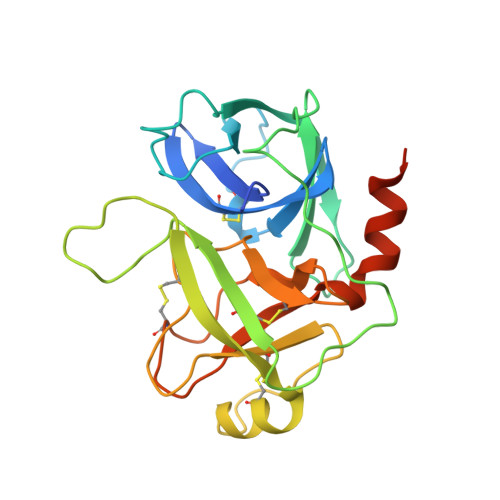
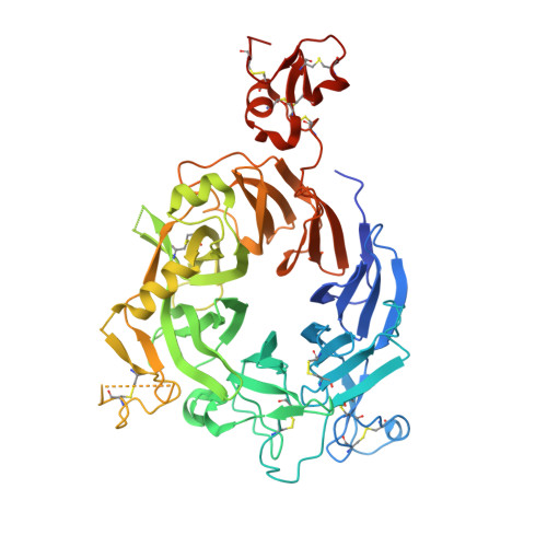
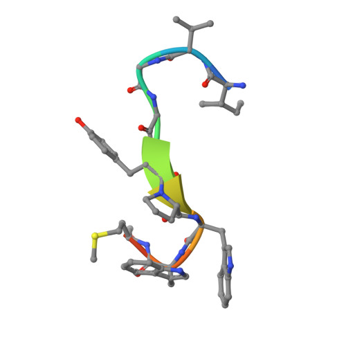
    Stimulation of hepatocyte growth factor (HGF) signaling through the Met receptor is an attractive approach for promoting tissue repair and preventing fibrosis. Using structure-guided peptide phage display combined with an activity-based sorting strategy, we engineered allosteric activators of zymogen-like pro-HGF to bypass proteolytic activation and reversibly stimulate pro-HGF signaling through Met. Biochemical, structural and biological data showed that zymogen activator peptides (ZAPtides) potently and selectively bind the activation pocket within the serine protease-like β-chain of pro-HGF and display titratable activation of pro-HGF-dependent Met signaling, leading to cell survival and migration. To further demonstrate the versatility of our ZAPtide platform, we identified allosteric activators for pro-macrophage stimulating protein and a zymogen serine protease, Protein C, which also provides evidence for target selectivity. These studies reveal that ZAPtides use molecular mimicry of the trypsin-like N-terminal insertion mechanism and establish a new paradigm for selective pharmacological activation of plasminogen-related growth factors and zymogen serine proteases.


  • Organizational Affiliation
    • 1] Department of Early Discovery Biochemistry, Genentech, Inc., San Francisco, California, USA. [2].

Macromolecule Content 

  • Total Structure Weight: 91.65 kDa 
  • Atom Count: 5,804 
  • Modeled Residue Count: 727 
  • Deposited Residue Count: 802 
  • Unique protein chains: 3

Macromolecules

Find similar proteins by:|  3D Structure
Entity ID: 1
MoleculeChains  Sequence LengthOrganismDetailsImage
Hepatocyte growth factor240Homo sapiensMutation(s): 2 
Gene Names: HGFHPTA
UniProt & NIH Common Fund Data Resources
Find proteins for P14210 (Homo sapiens)
Explore P14210 
Go to UniProtKB:  P14210
PHAROS:  P14210
GTEx:  ENSG00000019991 
Entity Groups
Sequence Clusters30% Identity50% Identity70% Identity90% Identity95% Identity100% Identity
UniProt GroupP14210
Sequence Annotations
Expand
Reference Sequence
Find similar proteins by:|  3D Structure
Entity ID: 2
MoleculeChains  Sequence LengthOrganismDetailsImage
Hepatocyte growth factor receptor551Homo sapiensMutation(s): 0 
Gene Names: MET
EC: 2.7.10.1
UniProt & NIH Common Fund Data Resources
Find proteins for P08581 (Homo sapiens)
Explore P08581 
Go to UniProtKB:  P08581
PHAROS:  P08581
GTEx:  ENSG00000105976 
Entity Groups
Sequence Clusters30% Identity50% Identity70% Identity90% Identity95% Identity100% Identity
UniProt GroupP08581
Glycosylation
Glycosylation Sites: 1Go to GlyGen: P08581-1
Sequence Annotations
Expand
Reference Sequence
Find similar proteins by:  Sequence   |   3D Structure  
Entity ID: 3
MoleculeChains  Sequence LengthOrganismDetailsImage
ZAP.14C [auth P]11synthetic constructMutation(s): 0 

Oligosaccharides

Help  
Entity ID: 4
MoleculeChains Length2D Diagram GlycosylationD Interactions
alpha-L-fucopyranose-(1-3)-[2-acetamido-2-deoxy-beta-D-glucopyranose-(1-4)]2-acetamido-2-deoxy-beta-D-glucopyranoseD [auth C]3N-Glycosylation
Glycosylation Resources
GlyTouCan: G11971MR
GlyCosmos: G11971MR
GlyGen: G11971MR

Small Molecules

Ligands 1 Unique
IDChains Name / Formula / InChI Key2D Diagram3D Interactions
1PE

Query on 1PE


Download:Ideal Coordinates CCD File
E [auth A]PENTAETHYLENE GLYCOL
C10 H22 O6
JLFNLZLINWHATN-UHFFFAOYSA-N

Experimental Data & Validation

Experimental Data

  • Method: X-RAY DIFFRACTION
  • Resolution: 2.99 Å
  • R-Value Free:  0.276 (Depositor), 0.293 (DCC) 
  • R-Value Work:  0.232 (Depositor), 0.243 (DCC) 
  • R-Value Observed: 0.233 (Depositor) 
Space Group: P 21 21 21
Unit Cell:
Length ( Å )Angle ( ˚ )
a = 59.789α = 90
b = 121.467β = 90
c = 137.657γ = 90
Software Package:
Software NamePurpose
MAR345dtbdata collection
PHASERphasing
BUSTERrefinement
HKL-2000data reduction
SCALEPACKdata scaling

Structure Validation

View Full Validation Report


Entry History 

Deposition Data

Revision History  (Full details and data files)

  • Version 1.0: 2014-06-04
    Type: Initial release
  • Version 1.1: 2014-07-02
    Changes: Database references
  • Version 2.0: 2020-07-29
    Type: Remediation
    Reason: Carbohydrate remediation
    Changes: Advisory, Atomic model, Data collection, Database references, Derived calculations, Structure summary
  • Version 2.1: 2023-09-20
    Changes: Data collection, Database references, Refinement description, Structure summary
  • Version 2.2: 2024-10-09
    Changes: Structure summary