4NST

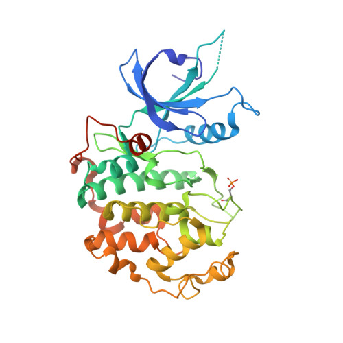
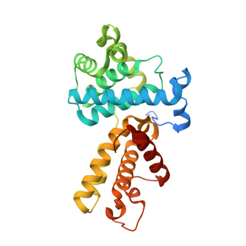
Crystal structure of human Cdk12/Cyclin K in complex with ADP-aluminum fluoride


Experimental Data Snapshot

  • Method: X-RAY DIFFRACTION
  • Resolution: 2.20 Å
  • R-Value Free: 0.240 
  • R-Value Work: 0.195 
  • R-Value Observed: 0.198 

Starting Models: experimental
View more details

wwPDB Validation   3D Report Full Report


Ligand Structure Quality Assessment 


This is version 1.2 of the entry. See complete history


Literature

The structure and substrate specificity of human Cdk12/Cyclin K.

Bosken, C.A.Farnung, L.Hintermair, C.Merzel Schachter, M.Vogel-Bachmayr, K.Blazek, D.Anand, K.Fisher, R.P.Eick, D.Geyer, M.

(2014) Nat Commun 5: 3505-3505

  • DOI: https://doi.org/10.1038/ncomms4505
  • Primary Citation of Related Structures:  
    4NST

  • PubMed Abstract: 

    Phosphorylation of the RNA polymerase II C-terminal domain (CTD) by cyclin-dependent kinases is important for productive transcription. Here we determine the crystal structure of Cdk12/CycK and analyse its requirements for substrate recognition. Active Cdk12/CycK is arranged in an open conformation similar to that of Cdk9/CycT but different from those of cell cycle kinases. Cdk12 contains a C-terminal extension that folds onto the N- and C-terminal lobes thereby contacting the ATP ribose. The interaction is mediated by an HE motif followed by a polybasic cluster that is conserved in transcriptional CDKs. Cdk12/CycK showed the highest activity on a CTD substrate prephosphorylated at position Ser7, whereas the common Lys7 substitution was not recognized. Flavopiridol is most potent towards Cdk12 but was still 10-fold more potent towards Cdk9. T-loop phosphorylation of Cdk12 required coexpression with a Cdk-activating kinase. These results suggest the regulation of Pol II elongation by a relay of transcriptionally active CTD kinases.


  • Organizational Affiliation

    1] Group Physical Biochemistry, Center of Advanced European Studies and Research, Ludwig-Erhard-Allee 2, Bonn 53175, Germany [2] Department of Physical Biochemistry, Max Planck Institute of Molecular Physiology, Otto-Hahn-Strasse 11, Dortmund 44227, Germany.


Macromolecules
Find similar proteins by:  (by identity cutoff)  |  3D Structure
Entity ID: 1
MoleculeChains Sequence LengthOrganismDetailsImage
Cyclin-dependent kinase 12
A, C
351Homo sapiensMutation(s): 0 
Gene Names: CDK12CRK7CRKRSKIAA0904
EC: 2.7.11.22 (PDB Primary Data), 2.7.11.23 (PDB Primary Data)
UniProt & NIH Common Fund Data Resources
Find proteins for Q9NYV4 (Homo sapiens)
Explore Q9NYV4 
Go to UniProtKB:  Q9NYV4
PHAROS:  Q9NYV4
GTEx:  ENSG00000167258 
Entity Groups  
Sequence Clusters30% Identity50% Identity70% Identity90% Identity95% Identity100% Identity
UniProt GroupQ9NYV4
Sequence Annotations
Expand
  • Reference Sequence
Find similar proteins by:  (by identity cutoff)  |  3D Structure
Entity ID: 2
MoleculeChains Sequence LengthOrganismDetailsImage
Cyclin-K
B, D
268Homo sapiensMutation(s): 0 
Gene Names: CCNKCPR4
UniProt & NIH Common Fund Data Resources
Find proteins for O75909 (Homo sapiens)
Explore O75909 
Go to UniProtKB:  O75909
PHAROS:  O75909
GTEx:  ENSG00000090061 
Entity Groups  
Sequence Clusters30% Identity50% Identity70% Identity90% Identity95% Identity100% Identity
UniProt GroupO75909
Sequence Annotations
Expand
  • Reference Sequence
Small Molecules
Ligands 4 Unique
IDChains Name / Formula / InChI Key2D Diagram3D Interactions
ADP
Query on ADP

Download Ideal Coordinates CCD File 
E [auth A],
L [auth C]
ADENOSINE-5'-DIPHOSPHATE
C10 H15 N5 O10 P2
XTWYTFMLZFPYCI-KQYNXXCUSA-N
AF3
Query on AF3

Download Ideal Coordinates CCD File 
F [auth A],
M [auth C]
ALUMINUM FLUORIDE
Al F3
KLZUFWVZNOTSEM-UHFFFAOYSA-K
EDO
Query on EDO

Download Ideal Coordinates CCD File 
J [auth A],
K [auth B],
P [auth C],
S [auth D]
1,2-ETHANEDIOL
C2 H6 O2
LYCAIKOWRPUZTN-UHFFFAOYSA-N
MG
Query on MG

Download Ideal Coordinates CCD File 
G [auth A]
H [auth A]
I [auth A]
N [auth C]
O [auth C]
G [auth A],
H [auth A],
I [auth A],
N [auth C],
O [auth C],
Q [auth D],
R [auth D]
MAGNESIUM ION
Mg
JLVVSXFLKOJNIY-UHFFFAOYSA-N
Modified Residues  1 Unique
IDChains TypeFormula2D DiagramParent
TPO
Query on TPO
A, C
L-PEPTIDE LINKINGC4 H10 N O6 PTHR
Experimental Data & Validation

Experimental Data

  • Method: X-RAY DIFFRACTION
  • Resolution: 2.20 Å
  • R-Value Free: 0.240 
  • R-Value Work: 0.195 
  • R-Value Observed: 0.198 
  • Space Group: P 1
Unit Cell:
Length ( Å )Angle ( ˚ )
a = 49.72α = 104.04
b = 78.85β = 85.76
c = 91.49γ = 101.46
Software Package:
Software NamePurpose
ADSCdata collection
PHASERphasing
PHENIXrefinement
XDSdata reduction
XSCALEdata scaling

Structure Validation

View Full Validation Report


Ligand Structure Quality Assessment 


Entry History 

Deposition Data

Revision History  (Full details and data files)

  • Version 1.0: 2014-03-26
    Type: Initial release
  • Version 1.1: 2014-10-01
    Changes: Database references
  • Version 1.2: 2023-09-20
    Changes: Data collection, Database references, Derived calculations, Refinement description