4NSD | pdb_00004nsd

Crystal Structure of CBARA1 in the Ca2+ Binding Form


Experimental Data Snapshot

  • Method: X-RAY DIFFRACTION
  • Resolution: 2.70 Å
  • R-Value Free: 
    0.282 (Depositor), 0.276 (DCC) 
  • R-Value Work: 
    0.212 (Depositor), 0.210 (DCC) 
  • R-Value Observed: 
    0.216 (Depositor) 

wwPDB Validation 3D Report Full Report

Validation slider image for 4NSD

This is version 1.2 of the entry. See complete history

Literature

Structural and mechanistic insights into MICU1 regulation of mitochondrial calcium uptake.

Wang, L.Yang, X.Li, S.Wang, Z.Liu, Y.Feng, J.Zhu, Y.Shen, Y.

(2014) EMBO J 33: 594-604

  • DOI: https://doi.org/10.1002/embj.201386523
  • Primary Citation Related Structures: 
    4NSC, 4NSD

  • PubMed Abstract: 

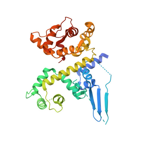
    Mitochondrial calcium uptake is a critical event in various cellular activities. Two recently identified proteins, the mitochondrial Ca(2+) uniporter (MCU), which is the pore-forming subunit of a Ca(2+) channel, and mitochondrial calcium uptake 1 (MICU1), which is the regulator of MCU, are essential in this event. However, the molecular mechanism by which MICU1 regulates MCU remains elusive. In this study, we report the crystal structures of Ca(2+)-free and Ca(2+)-bound human MICU1. Our studies reveal that Ca(2+)-free MICU1 forms a hexamer that binds and inhibits MCU. Upon Ca(2+) binding, MICU1 undergoes large conformational changes, resulting in the formation of multiple oligomers to activate MCU. Furthermore, we demonstrate that the affinity of MICU1 for Ca(2+) is approximately 15-20 μM. Collectively, our results provide valuable details to decipher the molecular mechanism of MICU1 regulation of mitochondrial calcium uptake.


  • Organizational Affiliation
    • State Key Laboratory of Medicinal Chemical Biology, Nankai University, Tianjin, China.

Macromolecule Content 

  • Total Structure Weight: 81.55 kDa 
  • Atom Count: 4,591 
  • Modeled Residue Count: 573 
  • Deposited Residue Count: 702 
  • Unique protein chains: 1

Macromolecules

Find similar proteins by:|  3D Structure
Entity ID: 1
MoleculeChains  Sequence LengthOrganismDetailsImage
Calcium uptake protein 1, mitochondrial
A, B
351Homo sapiensMutation(s): 0 
Gene Names: MICU1CALCCBARA1
UniProt & NIH Common Fund Data Resources
Find proteins for Q9BPX6 (Homo sapiens)
Explore Q9BPX6 
Go to UniProtKB:  Q9BPX6
PHAROS:  Q9BPX6
GTEx:  ENSG00000107745 
Entity Groups
Sequence Clusters30% Identity50% Identity70% Identity90% Identity95% Identity100% Identity
UniProt GroupQ9BPX6
Sequence Annotations
Expand
Reference Sequence

Small Molecules

Ligands 3 Unique
IDChains Name / Formula / InChI Key2D Diagram3D Interactions
MPD

Query on MPD


Download:Ideal Coordinates CCD File
H [auth B],
I [auth B],
J [auth B]
(4S)-2-METHYL-2,4-PENTANEDIOL
C6 H14 O2
SVTBMSDMJJWYQN-YFKPBYRVSA-N
CA

Query on CA


Download:Ideal Coordinates CCD File
C [auth A],
D [auth A],
F [auth B],
G [auth B]
CALCIUM ION
Ca
BHPQYMZQTOCNFJ-UHFFFAOYSA-N
CL

Query on CL


Download:Ideal Coordinates CCD File
E [auth A],
K [auth B]
CHLORIDE ION
Cl
VEXZGXHMUGYJMC-UHFFFAOYSA-M

Experimental Data & Validation

Experimental Data

  • Method: X-RAY DIFFRACTION
  • Resolution: 2.70 Å
  • R-Value Free:  0.282 (Depositor), 0.276 (DCC) 
  • R-Value Work:  0.212 (Depositor), 0.210 (DCC) 
  • R-Value Observed: 0.216 (Depositor) 
Space Group: I 21 21 21
Unit Cell:
Length ( Å )Angle ( ˚ )
a = 99.283α = 90
b = 105.804β = 90
c = 156.851γ = 90
Software Package:
Software NamePurpose
MAR345dtbdata collection
PHASERphasing
PHENIXrefinement
HKL-2000data reduction
HKL-2000data scaling

Structure Validation

View Full Validation Report


Entry History 

Deposition Data

Revision History  (Full details and data files)

  • Version 1.0: 2014-02-26
    Type: Initial release
  • Version 1.1: 2014-04-23
    Changes: Database references
  • Version 1.2: 2024-02-28
    Changes: Data collection, Database references, Derived calculations