4NNX | pdb_00004nnx

Crystal structure of PKD2 phosphopeptide bound to HLA-A2


Experimental Data Snapshot

  • Method: X-RAY DIFFRACTION
  • Resolution: 2.10 Å
  • R-Value Free: 
    0.236 (Depositor), 0.237 (DCC) 
  • R-Value Work: 
    0.198 (Depositor), 0.201 (DCC) 
  • R-Value Observed: 
    0.200 (Depositor) 

Starting Model: experimental
View more details

wwPDB Validation 3D Report Full Report

Validation slider image for 4NNX

This is version 1.4 of the entry. See complete history

Literature

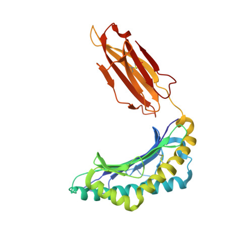
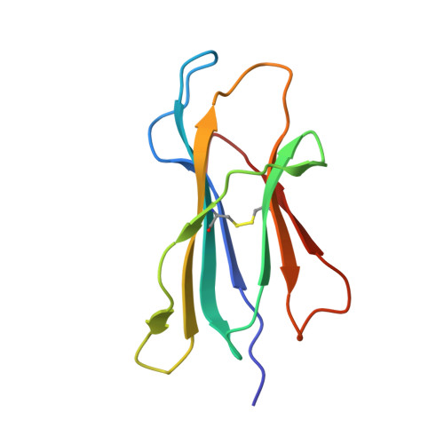
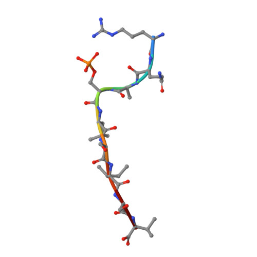
The antigenic identity of human class I MHC phosphopeptides is critically dependent upon phosphorylation status.

Mohammed, F.Stones, D.H.Zarling, A.L.Willcox, C.R.Shabanowitz, J.Cummings, K.L.Hunt, D.F.Cobbold, M.Engelhard, V.H.Willcox, B.E.

(2017) Oncotarget 8: 54160-54172

  • DOI: https://doi.org/10.18632/oncotarget.16952
  • Primary Citation Related Structures: 
    4NNX, 4NNY, 4NO0, 4NO2, 4NO3, 4NO5

  • PubMed Abstract: 

    Dysregulated post-translational modification provides a source of altered self-antigens that can stimulate immune responses in autoimmunity, inflammation, and cancer. In recent years, phosphorylated peptides have emerged as a group of tumour-associated antigens presented by MHC molecules and recognised by T cells, and represent promising candidates for cancer immunotherapy. However, the impact of phosphorylation on the antigenic identity of phosphopeptide epitopes is unclear. Here we examined this by determining structures of MHC-bound phosphopeptides bearing canonical position 4-phosphorylations in the presence and absence of their phosphate moiety, and examining phosphopeptide recognition by the T cell receptor (TCR). Strikingly, two peptides exhibited major conformational changes upon phosphorylation, involving a similar molecular mechanism, which focussed changes on the central peptide region most critical for T cell recognition. In contrast, a third epitope displayed little conformational alteration upon phosphorylation. In addition, binding studies demonstrated TCR interaction with an MHC-bound phosphopeptide was both epitope-specific and absolutely dependent upon phosphorylation status. These results highlight the critical influence of phosphorylation on the antigenic identity of naturally processed class I MHC epitopes. In doing so they provide a molecular framework for understanding phosphopeptide-specific immune responses, and have implications for the development of phosphopeptide antigen-specific cancer immunotherapy approaches.


  • Organizational Affiliation
    • Cancer Immunology and Immunotherapy Centre, Institute of Immunology and Immunotherapy, University of Birmingham, Edgbaston, Birmingham B15 2TT, UK.

Macromolecule Content 

  • Total Structure Weight: 45.06 kDa 
  • Atom Count: 3,353 
  • Modeled Residue Count: 382 
  • Deposited Residue Count: 382 
  • Unique protein chains: 3

Macromolecules

Find similar proteins by:|  3D Structure
Entity ID: 1
MoleculeChains  Sequence LengthOrganismDetailsImage
HLA class I histocompatibility antigen, A-2 alpha chain274Homo sapiensMutation(s): 0 
Gene Names: HLA-AHLAA
UniProt & NIH Common Fund Data Resources
Find proteins for P04439 (Homo sapiens)
Explore P04439 
Go to UniProtKB:  P04439
PHAROS:  P04439
GTEx:  ENSG00000206503 
Entity Groups
Sequence Clusters30% Identity50% Identity70% Identity90% Identity95% Identity100% Identity
UniProt GroupP04439
Sequence Annotations
Expand
Reference Sequence
Find similar proteins by:|  3D Structure
Entity ID: 2
MoleculeChains  Sequence LengthOrganismDetailsImage
Beta-2-microglobulin99Homo sapiensMutation(s): 0 
Gene Names: B2MCDABP0092HDCMA22P
UniProt & NIH Common Fund Data Resources
Find proteins for P61769 (Homo sapiens)
Explore P61769 
Go to UniProtKB:  P61769
PHAROS:  P61769
GTEx:  ENSG00000166710 
Entity Groups
Sequence Clusters30% Identity50% Identity70% Identity90% Identity95% Identity100% Identity
UniProt GroupP61769
Sequence Annotations
Expand
Reference Sequence
Find similar proteins by:  Sequence   |   3D Structure  
Entity ID: 3
MoleculeChains  Sequence LengthOrganismDetailsImage
Serine/threonine-protein kinase D29Homo sapiensMutation(s): 0 
EC: 2.7.11.13
UniProt & NIH Common Fund Data Resources
Find proteins for Q9BZL6 (Homo sapiens)
Explore Q9BZL6 
Go to UniProtKB:  Q9BZL6
PHAROS:  Q9BZL6
GTEx:  ENSG00000105287 
Entity Groups
UniProt GroupQ9BZL6
Sequence Annotations
Expand
Reference Sequence

Experimental Data & Validation

Experimental Data

  • Method: X-RAY DIFFRACTION
  • Resolution: 2.10 Å
  • R-Value Free:  0.236 (Depositor), 0.237 (DCC) 
  • R-Value Work:  0.198 (Depositor), 0.201 (DCC) 
  • R-Value Observed: 0.200 (Depositor) 
Space Group: P 21 21 21
Unit Cell:
Length ( Å )Angle ( ˚ )
a = 61.1α = 90
b = 79.8β = 90
c = 111.3γ = 90
Software Package:
Software NamePurpose
XSCALEdata scaling
MOLREPphasing
REFMACrefinement
PDB_EXTRACTdata extraction
CrystalCleardata collection

Structure Validation

View Full Validation Report


Entry History 

Deposition Data

Revision History  (Full details and data files)

  • Version 1.0: 2014-11-19
    Type: Initial release
  • Version 1.1: 2017-05-24
    Changes: Database references
  • Version 1.2: 2019-05-29
    Changes: Data collection, Database references, Derived calculations
  • Version 1.3: 2023-09-20
    Changes: Advisory, Data collection, Database references, Derived calculations, Refinement description
  • Version 1.4: 2024-10-09
    Changes: Structure summary