4NMQ | pdb_00004nmq

CFTR Associated Ligand (CAL) PDZ domain bound to peptide iCAL36(Ac-K-4) (ANSRW[Ac-K]TSII)


Experimental Data Snapshot

  • Method: X-RAY DIFFRACTION
  • Resolution: 1.40 Å
  • R-Value Free: 
    0.199 (Depositor), 0.199 (DCC) 
  • R-Value Work: 
    0.181 (Depositor), 0.178 (DCC) 
  • R-Value Observed: 
    0.182 (Depositor) 

Starting Model: experimental
View more details

wwPDB Validation 3D Report Full Report

Validation slider image for 4NMQ

This is version 1.3 of the entry. See complete history

Literature

Chemically Modified Peptide Scaffolds Target the CFTR-Associated Ligand PDZ Domain.

Amacher, J.F.Zhao, R.Spaller, M.R.Madden, D.R.

(2014) PLoS One 9: e103650-e103650

  • DOI: https://doi.org/10.1371/journal.pone.0103650
  • Primary Citation Related Structures: 
    4NMO, 4NMP, 4NMQ, 4NMR, 4NMS, 4NMT, 4NMV

  • PubMed Abstract: 

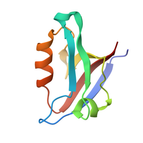
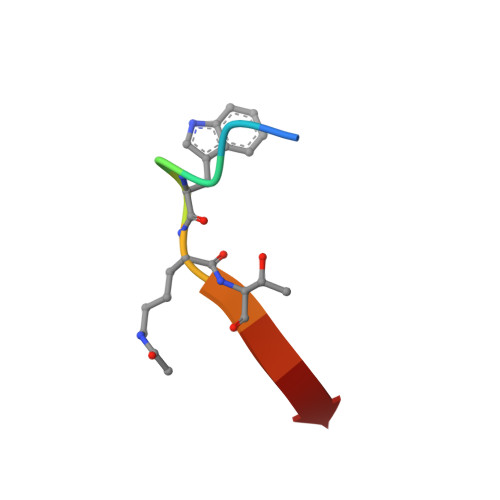
    PDZ domains are protein-protein interaction modules that coordinate multiple signaling and trafficking pathways in the cell and that include active therapeutic targets for diseases such as cancer, cystic fibrosis, and addiction. Our previous work characterized a PDZ interaction that restricts the apical membrane half-life of the cystic fibrosis transmembrane conductance regulator (CFTR). Using iterative cycles of peptide-array and solution-binding analysis, we targeted the PDZ domain of the CFTR-Associated Ligand (CAL), and showed that an engineered peptide inhibitor rescues cell-surface expression of the most common CFTR disease mutation ΔF508. Here, we present a series of scaffolds containing chemically modifiable side chains at all non-motif positions along the CAL PDZ domain binding cleft. Concordant equilibrium dissociation constants were determined in parallel by fluorescence polarization, isothermal titration calorimetry, and surface plasmon resonance techniques, confirming robust affinity for each scaffold and revealing an enthalpically driven mode of inhibitor binding. Structural studies demonstrate a conserved binding mode for each peptide, opening the possibility of combinatorial modification. Finally, we diversified one of our peptide scaffolds with halogenated substituents that yielded modest increases in binding affinity. Overall, this work validates our approach and provides a stereochemical foundation for further CAL inhibitor design and screening.


  • Organizational Affiliation
    • Department of Biochemistry, Geisel School of Medicine at Dartmouth, Hanover, New Hampshire, United States of America.

Macromolecule Content 

  • Total Structure Weight: 21.33 kDa 
  • Atom Count: 1,718 
  • Modeled Residue Count: 193 
  • Deposited Residue Count: 194 
  • Unique protein chains: 2

Macromolecules

Find similar proteins by:|  3D Structure
Entity ID: 1
MoleculeChains  Sequence LengthOrganismDetailsImage
Golgi-associated PDZ and coiled-coil motif-containing protein
A, B
87Homo sapiensMutation(s): 0 
Gene Names: GOPCCALFIG
UniProt & NIH Common Fund Data Resources
Find proteins for Q9HD26 (Homo sapiens)
Explore Q9HD26 
Go to UniProtKB:  Q9HD26
PHAROS:  Q9HD26
GTEx:  ENSG00000047932 
Entity Groups
Sequence Clusters30% Identity50% Identity70% Identity90% Identity95% Identity100% Identity
UniProt GroupQ9HD26
Sequence Annotations
Expand
Reference Sequence
Find similar proteins by:  Sequence   |   3D Structure  
Entity ID: 2
MoleculeChains  Sequence LengthOrganismDetailsImage
iCAL36(Ac-K-4) peptide
C, D
10N/AMutation(s): 0 

Small Molecules

Modified Residues  1 Unique
IDChains TypeFormula2D DiagramParent
ALY
Query on ALY
C, D
L-PEPTIDE LINKINGC8 H16 N2 O3LYS

Biologically Interesting Molecules (External Reference) 

1 Unique

Experimental Data & Validation

Experimental Data

  • Method: X-RAY DIFFRACTION
  • Resolution: 1.40 Å
  • R-Value Free:  0.199 (Depositor), 0.199 (DCC) 
  • R-Value Work:  0.181 (Depositor), 0.178 (DCC) 
  • R-Value Observed: 0.182 (Depositor) 
Space Group: P 21 21 21
Unit Cell:
Length ( Å )Angle ( ˚ )
a = 35.546α = 90
b = 47.462β = 90
c = 96.621γ = 90
Software Package:
Software NamePurpose
HKL-2000data collection
PHENIXmodel building
PHENIXrefinement
XDSdata reduction
XSCALEdata scaling
PHENIXphasing

Structure Validation

View Full Validation Report


Entry History 

Deposition Data

Revision History  (Full details and data files)

  • Version 1.0: 2014-10-01
    Type: Initial release
  • Version 1.1: 2023-09-20
    Changes: Data collection, Database references, Derived calculations, Refinement description
  • Version 1.2: 2023-12-06
    Changes: Data collection
  • Version 1.3: 2024-11-20
    Changes: Structure summary