4NDH

Human Aprataxin (Aptx) bound to DNA, AMP, and Zn - product complex


Experimental Data Snapshot

  • Method: X-RAY DIFFRACTION
  • Resolution: 1.85 Å
  • R-Value Free: 0.196 
  • R-Value Work: 0.163 
  • R-Value Observed: 0.164 

wwPDB Validation   3D Report Full Report


Ligand Structure Quality Assessment 


This is version 1.3 of the entry. See complete history


Literature

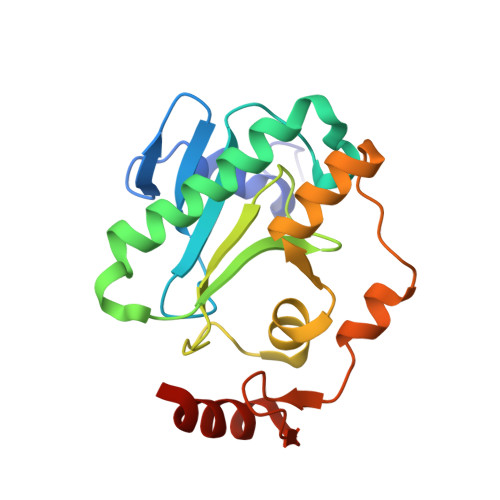
Aprataxin resolves adenylated RNA-DNA junctions to maintain genome integrity.

Tumbale, P.Williams, J.S.Schellenberg, M.J.Kunkel, T.A.Williams, R.S.

(2013) Nature 506: 111-115

  • DOI: https://doi.org/10.1038/nature12824
  • Primary Citation of Related Structures:  
    4NDF, 4NDG, 4NDH, 4NDI

  • PubMed Abstract: 

    Faithful maintenance and propagation of eukaryotic genomes is ensured by three-step DNA ligation reactions used by ATP-dependent DNA ligases. Paradoxically, when DNA ligases encounter nicked DNA structures with abnormal DNA termini, DNA ligase catalytic activity can generate and/or exacerbate DNA damage through abortive ligation that produces chemically adducted, toxic 5'-adenylated (5'-AMP) DNA lesions. Aprataxin (APTX) reverses DNA adenylation but the context for deadenylation repair is unclear. Here we examine the importance of APTX to RNase-H2-dependent excision repair (RER) of a lesion that is very frequently introduced into DNA, a ribonucleotide. We show that ligases generate adenylated 5' ends containing a ribose characteristic of RNase H2 incision. APTX efficiently repairs adenylated RNA-DNA, and acting in an RNA-DNA damage response (RDDR), promotes cellular survival and prevents S-phase checkpoint activation in budding yeast undergoing RER. Structure-function studies of human APTX-RNA-DNA-AMP-Zn complexes define a mechanism for detecting and reversing adenylation at RNA-DNA junctions. This involves A-form RNA binding, proper protein folding and conformational changes, all of which are affected by heritable APTX mutations in ataxia with oculomotor apraxia 1. Together, these results indicate that accumulation of adenylated RNA-DNA may contribute to neurological disease.


  • Organizational Affiliation

    1] Laboratory of Structural Biology, National Institute of Environmental Health Sciences, NIH, DHHS, Research Triangle Park, North Carolina 27709, USA [2].


Macromolecules

Find similar proteins by:  (by identity cutoff)  |  3D Structure
Entity ID: 1
MoleculeChains Sequence LengthOrganismDetailsImage
Aprataxin
A, B
182Homo sapiensMutation(s): 0 
Gene Names: APTXAXA1
UniProt & NIH Common Fund Data Resources
Find proteins for Q7Z2E3 (Homo sapiens)
Explore Q7Z2E3 
Go to UniProtKB:  Q7Z2E3
PHAROS:  Q7Z2E3
GTEx:  ENSG00000137074 
Entity Groups  
Sequence Clusters30% Identity50% Identity70% Identity90% Identity95% Identity100% Identity
UniProt GroupQ7Z2E3
Sequence Annotations
Expand
  • Reference Sequence

Find similar nucleic acids by:  Sequence   |   3D Structure  

Entity ID: 2
MoleculeChains LengthOrganismImage
5'-D(P*GP*TP*TP*CP*TP*AP*GP*AP*AP*C)-3'C [auth D],
D [auth E],
E [auth G],
F [auth H]
10N/A
Sequence Annotations
Expand
  • Reference Sequence
Experimental Data & Validation

Experimental Data

  • Method: X-RAY DIFFRACTION
  • Resolution: 1.85 Å
  • R-Value Free: 0.196 
  • R-Value Work: 0.163 
  • R-Value Observed: 0.164 
  • Space Group: P 21 21 21
Unit Cell:
Length ( Å )Angle ( ˚ )
a = 40.397α = 90
b = 117.082β = 90
c = 118.202γ = 90
Software Package:
Software NamePurpose
DENZOdata reduction
SCALEPACKdata scaling
PHENIXrefinement
PDB_EXTRACTdata extraction
SERGUIdata collection
PHASERphasing

Structure Validation

View Full Validation Report


Ligand Structure Quality Assessment 


Entry History 

Deposition Data

Revision History  (Full details and data files)

  • Version 1.0: 2013-12-18
    Type: Initial release
  • Version 1.1: 2014-01-15
    Changes: Database references
  • Version 1.2: 2014-02-05
    Changes: Database references
  • Version 1.3: 2023-09-20
    Changes: Data collection, Database references, Derived calculations, Refinement description