4NAM | pdb_00004nam

1.7A structure of 5-Fluoro Tryptophan Labeled Protective Antigen (W206Y)


Experimental Data Snapshot

  • Method: X-RAY DIFFRACTION
  • Resolution: 1.70 Å
  • R-Value Free: 
    0.208 (Depositor), 0.218 (DCC) 
  • R-Value Work: 
    0.179 (Depositor), 0.188 (DCC) 
  • R-Value Observed: 
    0.181 (Depositor) 

Starting Model: experimental
View more details

wwPDB Validation 3D Report Full Report

Validation slider image for 4NAM

This is version 1.3 of the entry. See complete history

Literature

(19)F nuclear magnetic resonance and crystallographic studies of 5-fluorotryptophan-labeled anthrax protective antigen and effects of the receptor on stability.

Chadegani, F.Lovell, S.Mullangi, V.Miyagi, M.Battaile, K.P.Bann, J.G.

(2014) Biochemistry 53: 690-701

  • DOI: https://doi.org/10.1021/bi401405s
  • Primary Citation Related Structures: 
    4NAM

  • PubMed Abstract: 

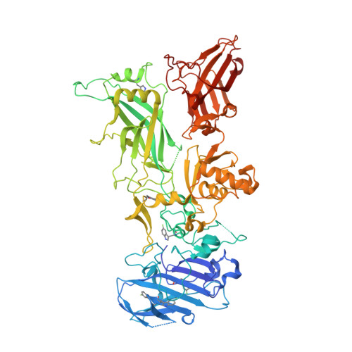
    The anthrax protective antigen (PA) is an 83 kDa protein that is one of three protein components of the anthrax toxin, an AB toxin secreted by Bacillus anthracis. PA is capable of undergoing several structural changes, including oligomerization to either a heptameric or octameric structure called the prepore, and at acidic pH a major conformational change to form a membrane-spanning pore. To follow these structural changes at a residue-specific level, we have conducted initial studies in which we have biosynthetically incorporated 5-fluorotryptophan (5-FTrp) into PA, and we have studied the influence of 5-FTrp labeling on the structural stability of PA and on binding to the host receptor capillary morphogenesis protein 2 (CMG2) using (19)F nuclear magnetic resonance (NMR). There are seven tryptophans in PA, but of the four domains in PA, only two contain tryptophans: domain 1 (Trp65, -90, -136, -206, and -226) and domain 2 (Trp346 and -477). Trp346 is of particular interest because of its proximity to the CMG2 binding interface, and because it forms part of the membrane-spanning pore. We show that the (19)F resonance of Trp346 is sensitive to changes in pH, consistent with crystallographic studies, and that receptor binding significantly stabilizes Trp346 to both pH and temperature. In addition, we provide evidence that suggests that resonances from tryptophans distant from the binding interface are also stabilized by the receptor. Our studies highlight the positive impact of receptor binding on protein stability and the use of (19)F NMR in gaining insight into structural changes in a high-molecular weight protein.


  • Organizational Affiliation
    • Department of Chemistry, Wichita State University , Wichita, Kansas 67260, United States.

Macromolecule Content 

  • Total Structure Weight: 82.93 kDa 
  • Atom Count: 5,753 
  • Modeled Residue Count: 682 
  • Deposited Residue Count: 735 
  • Unique protein chains: 1

Macromolecules

Find similar proteins by:|  3D Structure
Entity ID: 1
MoleculeChains  Sequence LengthOrganismDetailsImage
Protective antigen735Bacillus anthracisMutation(s): 1 
Gene Names: pagApagpXO1-110BXA0164GBAA_pXO1_0164
UniProt
Find proteins for P13423 (Bacillus anthracis)
Explore P13423 
Go to UniProtKB:  P13423
Entity Groups
Sequence Clusters30% Identity50% Identity70% Identity90% Identity95% Identity100% Identity
UniProt GroupP13423
Sequence Annotations
Expand
Reference Sequence

Small Molecules

Modified Residues  1 Unique
IDChains TypeFormula2D DiagramParent
FTR
Query on FTR
A
L-PEPTIDE LINKINGC11 H11 F N2 O2TRP

Experimental Data & Validation

Experimental Data

  • Method: X-RAY DIFFRACTION
  • Resolution: 1.70 Å
  • R-Value Free:  0.208 (Depositor), 0.218 (DCC) 
  • R-Value Work:  0.179 (Depositor), 0.188 (DCC) 
  • R-Value Observed: 0.181 (Depositor) 
Space Group: P 21 21 21
Unit Cell:
Length ( Å )Angle ( ˚ )
a = 71.298α = 90
b = 93.954β = 90
c = 117.727γ = 90
Software Package:
Software NamePurpose
SCALAdata scaling
PHASERphasing
PHENIXrefinement
PDB_EXTRACTdata extraction
JDirectordata collection
XDSdata reduction

Structure Validation

View Full Validation Report


Entry History 

Deposition Data

Revision History  (Full details and data files)

  • Version 1.0: 2014-01-22
    Type: Initial release
  • Version 1.1: 2014-12-24
    Changes: Database references
  • Version 1.2: 2017-11-15
    Changes: Refinement description
  • Version 1.3: 2023-09-20
    Changes: Data collection, Database references, Derived calculations, Refinement description