4MR1 | pdb_00004mr1

X-ray structure of the adduct between hen egg white lysozyme and cis-diamminediiodoplatinum(II)


Experimental Data Snapshot

  • Method: X-RAY DIFFRACTION
  • Resolution: 1.99 Å
  • R-Value Free: 
    0.234 (Depositor), 0.242 (DCC) 
  • R-Value Work: 
    0.184 (Depositor), 0.196 (DCC) 
  • R-Value Observed: 
    0.184 (Depositor) 

wwPDB Validation   3D Report Full Report


This is version 1.1 of the entry. See complete history


Literature

Peculiar features in the crystal structure of the adduct formed between cis-PtI2(NH3)2 and hen egg white lysozyme.

Messori, L.Marzo, T.Gabbiani, C.Valdes, A.A.Quiroga, A.G.Merlino, A.

(2013) Inorg Chem 52: 13827-13829

  • DOI: https://doi.org/10.1021/ic402611m
  • Primary Citation of Related Structures:  
    4MR1

  • PubMed Abstract: 

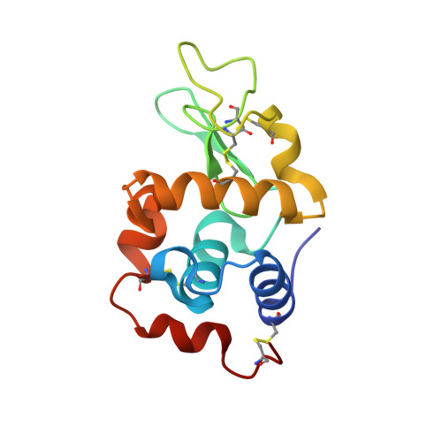
    The reactivity of cis-diamminediiodidoplatinum(II), cis-PtI2(NH3)2, the iodo analogue of cisplatin, with hen egg white lysozyme (HEWL) was investigated by electrospray ionization mass spectrometry and X-ray crystallography. Interestingly, the study compound forms a stable 1:1 protein adduct for which the crystal structure was solved at 1.99 Å resolution. In this adduct, the Pt(II) center, upon release of one ammonia ligand, selectively coordinates to the imidazole of His15. Both iodide ligands remain bound to platinum, with this being a highly peculiar and unexpected feature. Notably, two equivalent modes of Pt(II) binding are possible that differ only in the location of I atoms with respect to ND1 of His15. The structure of the adduct was compared with that of HEWL-cisplatin, previously described; differences are stressed and their important mechanistic implications discussed.


  • Organizational Affiliation
    • Department of Chemistry, University of Florence , Via della Lastruccia 3, 50019 Sesto Fiorentino, Italy.

Macromolecules
Find similar proteins by:  (by identity cutoff)  |  3D Structure
Entity ID: 1
MoleculeChains Sequence LengthOrganismDetailsImage
Lysozyme C129Gallus gallusMutation(s): 0 
EC: 3.2.1.17
UniProt
Find proteins for P00698 (Gallus gallus)
Explore P00698 
Go to UniProtKB:  P00698
Entity Groups  
Sequence Clusters30% Identity50% Identity70% Identity90% Identity95% Identity100% Identity
UniProt GroupP00698
Sequence Annotations
Expand
  • Reference Sequence
Experimental Data & Validation

Experimental Data

  • Method: X-RAY DIFFRACTION
  • Resolution: 1.99 Å
  • R-Value Free:  0.234 (Depositor), 0.242 (DCC) 
  • R-Value Work:  0.184 (Depositor), 0.196 (DCC) 
  • R-Value Observed: 0.184 (Depositor) 
Space Group: P 43 21 2
Unit Cell:
Length ( Å )Angle ( ˚ )
a = 77.017α = 90
b = 77.017β = 90
c = 36.834γ = 90
Software Package:
Software NamePurpose
CrystalCleardata collection
AMoREphasing
CNSrefinement
HKL-2000data reduction
HKL-2000data scaling

Structure Validation

View Full Validation Report


Entry History 

Deposition Data

  • Released Date: 2014-06-04 
  • Deposition Author(s): Merlino, A.

Revision History  (Full details and data files)

  • Version 1.0: 2014-06-04
    Type: Initial release
  • Version 1.1: 2024-11-20
    Changes: Data collection, Database references, Derived calculations, Structure summary