4MMZ | pdb_00004mmz

Integrin AlphaVBeta3 ectodomain bound to an antagonistic tenth domain of Fibronectin


Experimental Data Snapshot

  • Method: X-RAY DIFFRACTION
  • Resolution: 3.10 Å
  • R-Value Free: 
    0.255 (Depositor), 0.255 (DCC) 
  • R-Value Work: 
    0.207 (Depositor), 0.207 (DCC) 
  • R-Value Observed: 
    0.209 (Depositor) 

Starting Model: experimental
View more details

wwPDB Validation 3D Report Full Report

Validation slider image for 4MMZ

Ligand Structure Quality Assessment 


This is version 3.2 of the entry. See complete history

Literature

Structural basis for pure antagonism of integrin alpha V beta 3 by a high-affinity form of fibronectin.

Van Agthoven, J.F.Xiong, J.P.Alonso, J.L.Rui, X.Adair, B.D.Goodman, S.L.Arnaout, M.A.

(2014) Nat Struct Mol Biol 21: 383-388

  • DOI: https://doi.org/10.1038/nsmb.2797
  • Primary Citation Related Structures: 
    4MMX, 4MMY, 4MMZ

  • PubMed Abstract: 

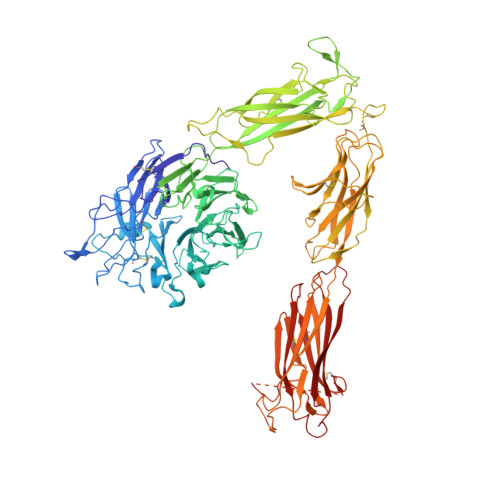
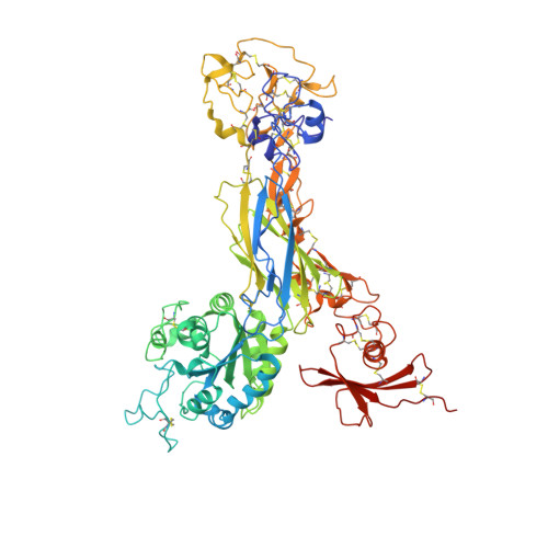
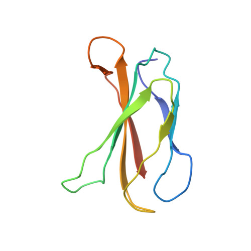
    Integrins are important therapeutic targets. However, current RGD-based anti-integrin drugs are also partial agonists, inducing conformational changes that trigger potentially fatal immune reactions and paradoxical cell adhesion. Here we describe the first crystal structure of αVβ3 bound to a physiologic ligand, the tenth type III RGD domain of wild-type fibronectin (wtFN10), or to a high-affinity mutant (hFN10) shown here to act as a pure antagonist. Comparison of these structures revealed a central π-π interaction between Trp1496 in the RGD-containing loop of hFN10 and Tyr122 of the β3 subunit that blocked conformational changes triggered by wtFN10 and trapped hFN10-bound αVβ3 in an inactive conformation. Removing the Trp1496 or Tyr122 side chains or reorienting Trp1496 away from Tyr122 converted hFN10 into a partial agonist. These findings offer new insights into the mechanism of integrin activation and a basis for the design of RGD-based pure antagonists.


  • Organizational Affiliation
    • 1] Structural Biology Program, Department of Medicine, Harvard Medical School, Massachusetts General Hospital, Charlestown, Massachusetts, USA. [2].

Macromolecule Content 

  • Total Structure Weight: 198.88 kDa 
  • Atom Count: 13,512 
  • Modeled Residue Count: 1,701 
  • Deposited Residue Count: 1,749 
  • Unique protein chains: 3

Macromolecules

Find similar proteins by:|  3D Structure
Entity ID: 1
MoleculeChains  Sequence LengthOrganismDetailsImage
Integrin alpha-V959Homo sapiensMutation(s): 0 
Gene Names: ITGAVMSK8VNRAVTNR
UniProt & NIH Common Fund Data Resources
Find proteins for P06756 (Homo sapiens)
Explore P06756 
Go to UniProtKB:  P06756
PHAROS:  P06756
GTEx:  ENSG00000138448 
Entity Groups
Sequence Clusters30% Identity50% Identity70% Identity90% Identity95% Identity100% Identity
UniProt GroupP06756
Glycosylation
Glycosylation Sites: 9Go to GlyGen: P06756-1
Sequence Annotations
Expand
Reference Sequence
Find similar proteins by:|  3D Structure
Entity ID: 2
MoleculeChains  Sequence LengthOrganismDetailsImage
Integrin beta-3692Homo sapiensMutation(s): 0 
Gene Names: ITGB3GP3A
UniProt & NIH Common Fund Data Resources
Find proteins for P05106 (Homo sapiens)
Explore P05106 
Go to UniProtKB:  P05106
PHAROS:  P05106
GTEx:  ENSG00000259207 
Entity Groups
Sequence Clusters30% Identity50% Identity70% Identity90% Identity95% Identity100% Identity
UniProt GroupP05106
Glycosylation
Glycosylation Sites: 5Go to GlyGen: P05106-1
Sequence Annotations
Expand
Reference Sequence
Find similar proteins by:|  3D Structure
Entity ID: 3
MoleculeChains  Sequence LengthOrganismDetailsImage
Fibronectin98Homo sapiensMutation(s): 5 
Gene Names: FN1FN
UniProt & NIH Common Fund Data Resources
Find proteins for P02751 (Homo sapiens)
Explore P02751 
Go to UniProtKB:  P02751
PHAROS:  P02751
GTEx:  ENSG00000115414 
Entity Groups
Sequence Clusters30% Identity50% Identity70% Identity90% Identity95% Identity100% Identity
UniProt GroupP02751
Sequence Annotations
Expand
Reference Sequence

Oligosaccharides

Help  
Entity ID: 4
MoleculeChains Length2D Diagram GlycosylationD Interactions
2-acetamido-2-deoxy-beta-D-glucopyranose-(1-4)-2-acetamido-2-deoxy-beta-D-glucopyranose
D, F, G, H
2N-Glycosylation
Glycosylation Resources
GlyTouCan: G42666HT
GlyCosmos: G42666HT
GlyGen: G42666HT
Entity ID: 5
MoleculeChains Length2D Diagram GlycosylationD Interactions
alpha-D-mannopyranose-(1-4)-beta-D-mannopyranose-(1-6)-[alpha-D-mannopyranose-(1-3)]beta-D-mannopyranose-(1-4)-2-acetamido-2-deoxy-beta-D-glucopyranose-(1-4)-2-acetamido-2-deoxy-beta-D-glucopyranose
E
6N-Glycosylation
Glycosylation Resources
GlyTouCan: G93290PO
GlyCosmos: G93290PO
GlyGen: G93290PO
Entity ID: 6
MoleculeChains Length2D Diagram GlycosylationD Interactions
beta-D-mannopyranose-(1-4)-2-acetamido-2-deoxy-beta-D-glucopyranose-(1-4)-2-acetamido-2-deoxy-beta-D-glucopyranose
I
3N-Glycosylation
Glycosylation Resources
GlyTouCan: G15407YE
GlyCosmos: G15407YE
GlyGen: G15407YE

Small Molecules

Ligands 4 Unique
IDChains Name / Formula / InChI Key2D Diagram3D Interactions
NAG

Query on NAG


Download:Ideal Coordinates CCD File
J [auth A]
K [auth A]
L [auth A]
M [auth A]
N [auth A]
J [auth A],
K [auth A],
L [auth A],
M [auth A],
N [auth A],
V [auth B],
W [auth B],
X [auth B]
2-acetamido-2-deoxy-beta-D-glucopyranose
C8 H15 N O6
OVRNDRQMDRJTHS-FMDGEEDCSA-N
GOL

Query on GOL


Download:Ideal Coordinates CCD File
BA [auth C],
U [auth A]
GLYCEROL
C3 H8 O3
PEDCQBHIVMGVHV-UHFFFAOYSA-N
MN

Query on MN


Download:Ideal Coordinates CCD File
AA [auth B]
O [auth A]
P [auth A]
Q [auth A]
R [auth A]
AA [auth B],
O [auth A],
P [auth A],
Q [auth A],
R [auth A],
S [auth A],
Y [auth B],
Z [auth B]
MANGANESE (II) ION
Mn
WAEMQWOKJMHJLA-UHFFFAOYSA-N
NA

Query on NA


Download:Ideal Coordinates CCD File
T [auth A]SODIUM ION
Na
FKNQFGJONOIPTF-UHFFFAOYSA-N

Experimental Data & Validation

Experimental Data

  • Method: X-RAY DIFFRACTION
  • Resolution: 3.10 Å
  • R-Value Free:  0.255 (Depositor), 0.255 (DCC) 
  • R-Value Work:  0.207 (Depositor), 0.207 (DCC) 
  • R-Value Observed: 0.209 (Depositor) 
Space Group: P 32 2 1
Unit Cell:
Length ( Å )Angle ( ˚ )
a = 129.792α = 90
b = 129.792β = 90
c = 307.675γ = 120
Software Package:
Software NamePurpose
DENZOdata reduction
SCALEPACKdata scaling
PHASERphasing
PHENIXrefinement
HKL-2000data reduction
HKL-2000data scaling

Structure Validation

View Full Validation Report


Ligand Structure Quality Assessment 


Entry History 

& Funding Information

Deposition Data


Funding OrganizationLocationGrant Number
National Institutes of Health/National Institute of Diabetes and Digestive and Kidney Disease (NIH/NIDDK)United States--

Revision History  (Full details and data files)

  • Version 1.0: 2014-03-26
    Type: Initial release
  • Version 1.1: 2014-04-09
    Changes: Database references
  • Version 1.2: 2014-04-30
    Changes: Database references
  • Version 1.3: 2017-11-15
    Changes: Refinement description
  • Version 2.0: 2020-07-29
    Type: Remediation
    Reason: Carbohydrate remediation
    Changes: Atomic model, Data collection, Database references, Derived calculations, Structure summary
  • Version 3.0: 2022-12-21
    Type: Coordinate replacement
    Reason: Atomic clashes
    Changes: Atomic model, Author supporting evidence, Data collection, Database references, Derived calculations, Non-polymer description, Other, Refinement description, Source and taxonomy, Structure summary
  • Version 3.1: 2023-09-20
    Changes: Data collection, Refinement description
  • Version 3.2: 2024-11-20
    Changes: Structure summary