4MLH

Human Glucokinase in Complex with a Novel Amino Thiazole Allosteric Activator


Experimental Data Snapshot

  • Method: X-RAY DIFFRACTION
  • Resolution: 2.90 Å
  • R-Value Free: 0.282 
  • R-Value Work: 0.222 

wwPDB Validation   3D Report Full Report


Ligand Structure Quality Assessment 


This is version 1.3 of the entry. See complete history


Literature

Identification of a New Class of Glucokinase Activators through Structure-Based Design.

Hinklin, R.J.Boyd, S.A.Chicarelli, M.J.Condroski, K.R.Dewolf, W.E.Lee, P.A.Lee, W.Singh, A.Thomas, L.Voegtli, W.C.Williams, L.Aicher, T.D.

(2013) J Med Chem 56: 7669-7678

  • DOI: https://doi.org/10.1021/jm401116k
  • Primary Citation of Related Structures:  
    4MLE, 4MLH

  • PubMed Abstract: 

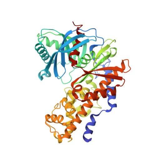
    Glucose flux through glucokinase (GK) controls insulin release from the pancreas in response to high glucose concentrations. Glucose flux through GK also contributes to reducing hepatic glucose output. Because many individuals with type 2 diabetes appear to have an inadequacy or defect in one or both of these processes, compounds that can activate GK may serve as effective treatments for type 2 diabetes. Herein we report the identification and initial optimization of a novel series of allosteric glucokinase activators (GKAs). We discovered an initial thiazolylamino pyridine-based hit that was optimized using a structure-based design strategy and identified 26 as an early lead. Compound 26 demonstrated a good balance of in vitro potency and enzyme kinetic parameters and demonstrated blood glucose reductions in oral glucose tolerance tests in both C57BL/6J mice and high-fat fed Zucker diabetic fatty rats.


  • Organizational Affiliation

    Array BioPharma , 3200 Walnut Street, Boulder, Colorado 80301, United States.


Macromolecules
Find similar proteins by:  (by identity cutoff)  |  3D Structure
Entity ID: 1
MoleculeChains Sequence LengthOrganismDetailsImage
Glucokinase455Homo sapiensMutation(s): 0 
Gene Names: GCK
EC: 2.7.1.2
UniProt & NIH Common Fund Data Resources
Find proteins for P35557 (Homo sapiens)
Explore P35557 
Go to UniProtKB:  P35557
PHAROS:  P35557
GTEx:  ENSG00000106633 
Entity Groups  
Sequence Clusters30% Identity50% Identity70% Identity90% Identity95% Identity100% Identity
UniProt GroupP35557
Sequence Annotations
Expand
  • Reference Sequence
Small Molecules
Ligands 2 Unique
IDChains Name / Formula / InChI Key2D Diagram3D Interactions
VO2
Query on VO2

Download Ideal Coordinates CCD File 
C [auth A]3-(benzyloxy)-5-methyl-N-(4-methyl-1,3-thiazol-2-yl)pyridin-2-amine
C17 H17 N3 O S
QFOFZZJJRFMLKB-UHFFFAOYSA-N
GLC
Query on GLC

Download Ideal Coordinates CCD File 
B [auth A]alpha-D-glucopyranose
C6 H12 O6
WQZGKKKJIJFFOK-DVKNGEFBSA-N
Binding Affinity Annotations 
IDSourceBinding Affinity
VO2 BindingDB:  4MLH EC50: 2640 (nM) from 1 assay(s)
Experimental Data & Validation

Experimental Data

  • Method: X-RAY DIFFRACTION
  • Resolution: 2.90 Å
  • R-Value Free: 0.282 
  • R-Value Work: 0.222 
  • Space Group: P 65 2 2
Unit Cell:
Length ( Å )Angle ( ˚ )
a = 78.77α = 90
b = 78.77β = 90
c = 319.62γ = 120
Software Package:
Software NamePurpose
CrystalCleardata collection
CNSrefinement
MOSFLMdata reduction
SCALAdata scaling
CNSphasing

Structure Validation

View Full Validation Report


Ligand Structure Quality Assessment 


Entry History 

Deposition Data

Revision History  (Full details and data files)

  • Version 1.0: 2013-09-25
    Type: Initial release
  • Version 1.1: 2013-10-23
    Changes: Database references
  • Version 1.2: 2020-07-29
    Type: Remediation
    Reason: Carbohydrate remediation
    Changes: Data collection, Database references, Derived calculations, Structure summary
  • Version 1.3: 2024-02-28
    Changes: Data collection, Database references, Structure summary