4ML5

one minute iron loaded frog M ferritin mutant H54Q


Experimental Data Snapshot

  • Method: X-RAY DIFFRACTION
  • Resolution: 1.22 Å
  • R-Value Free: 0.154 
  • R-Value Work: 0.139 
  • R-Value Observed: 0.140 

wwPDB Validation   3D Report Full Report


This is version 1.3 of the entry. See complete history


Literature

Time-lapse anomalous X-ray diffraction shows how Fe(2+) substrate ions move through ferritin protein nanocages to oxidoreductase sites.

Pozzi, C.Di Pisa, F.Lalli, D.Rosa, C.Theil, E.Turano, P.Mangani, S.

(2015) Acta Crystallogr D Biol Crystallogr 71: 941-953

  • DOI: https://doi.org/10.1107/S1399004715002333
  • Primary Citation of Related Structures:  
    4LPJ, 4LQH, 4LQJ, 4LQV, 4LYU, 4LYX, 4MJY, 4MKU, 4ML5, 4MN9, 4MY7, 6I36

  • PubMed Abstract: 

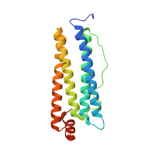
    Ferritin superfamily protein cages reversibly synthesize internal biominerals, Fe2O3·H2O. Fe(2+) and O2 (or H2O2) substrates bind at oxidoreductase sites in the cage, initiating biomineral synthesis to concentrate iron and prevent potentially toxic reactions products from Fe(2+)and O2 or H2O2 chemistry. By freezing ferritin crystals of Rana catesbeiana ferritin M (RcMf) at different time intervals after exposure to a ferrous salt, a series of high-resolution anomalous X-ray diffraction data sets were obtained that led to crystal structures that allowed the direct observation of ferrous ions entering, moving along and binding at enzyme sites in the protein cages. The ensemble of crystal structures from both aerobic and anaerobic conditions provides snapshots of the iron substrate bound at different cage locations that vary with time. The observed differential occupation of the two iron sites in the enzyme oxidoreductase centre (with Glu23 and Glu58, and with Glu58, His61 and Glu103 as ligands, respectively) and other iron-binding sites (with Glu53, His54, Glu57, Glu136 and Asp140 as ligands) reflects the approach of the Fe(2+) substrate and its progression before the enzymatic cycle 2Fe(2+) + O2 → Fe(3+)-O-O-Fe(3+) → Fe(3+)-O(H)-Fe(3+) and turnover. The crystal structures also revealed different Fe(2+) coordination compounds bound to the ion channels located at the threefold and fourfold symmetry axes of the cage.


  • Organizational Affiliation

    Dipartimento di Biotecnologie, Chimica e Farmacia, University of Siena, Via Aldo Moro 2, 53100 Siena, Italy.


Macromolecules
Find similar proteins by:  (by identity cutoff)  |  3D Structure
Entity ID: 1
MoleculeChains Sequence LengthOrganismDetailsImage
Ferritin, middle subunit176Aquarana catesbeianaMutation(s): 1 
EC: 1.16.3.1
UniProt
Find proteins for P07798 (Aquarana catesbeiana)
Explore P07798 
Go to UniProtKB:  P07798
Entity Groups  
Sequence Clusters30% Identity50% Identity70% Identity90% Identity95% Identity100% Identity
UniProt GroupP07798
Sequence Annotations
Expand
  • Reference Sequence
Small Molecules
Ligands 3 Unique
IDChains Name / Formula / InChI Key2D Diagram3D Interactions
FE2
Query on FE2

Download Ideal Coordinates CCD File 
B [auth A],
C [auth A],
D [auth A],
E [auth A]
FE (II) ION
Fe
CWYNVVGOOAEACU-UHFFFAOYSA-N
CL
Query on CL

Download Ideal Coordinates CCD File 
AA [auth A]
BA [auth A]
CA [auth A]
DA [auth A]
O [auth A]
AA [auth A],
BA [auth A],
CA [auth A],
DA [auth A],
O [auth A],
P [auth A],
Q [auth A],
R [auth A],
S [auth A],
T [auth A],
U [auth A],
V [auth A],
W [auth A],
X [auth A],
Y [auth A],
Z [auth A]
CHLORIDE ION
Cl
VEXZGXHMUGYJMC-UHFFFAOYSA-M
MG
Query on MG

Download Ideal Coordinates CCD File 
F [auth A]
G [auth A]
H [auth A]
I [auth A]
J [auth A]
F [auth A],
G [auth A],
H [auth A],
I [auth A],
J [auth A],
K [auth A],
L [auth A],
M [auth A],
N [auth A]
MAGNESIUM ION
Mg
JLVVSXFLKOJNIY-UHFFFAOYSA-N
Experimental Data & Validation

Experimental Data

  • Method: X-RAY DIFFRACTION
  • Resolution: 1.22 Å
  • R-Value Free: 0.154 
  • R-Value Work: 0.139 
  • R-Value Observed: 0.140 
  • Space Group: F 4 3 2
Unit Cell:
Length ( Å )Angle ( ˚ )
a = 184.23α = 90
b = 184.23β = 90
c = 184.23γ = 90
Software Package:
Software NamePurpose
MOLREPphasing
REFMACrefinement
MOSFLMdata reduction
SCALAdata scaling

Structure Validation

View Full Validation Report


Entry History 

Deposition Data

Revision History  (Full details and data files)

  • Version 1.0: 2014-09-10
    Type: Initial release
  • Version 1.1: 2014-10-01
    Changes: Non-polymer description
  • Version 1.2: 2015-04-15
    Changes: Database references
  • Version 1.3: 2023-09-20
    Changes: Data collection, Database references, Derived calculations, Refinement description