4MKY

Polymerase Domain from Mycobacterium tuberculosis Ligase D in complex with an annealed double-strand DNA break.


Experimental Data Snapshot

  • Method: X-RAY DIFFRACTION
  • Resolution: 2.40 Å
  • R-Value Free: 0.245 
  • R-Value Work: 0.197 
  • R-Value Observed: 0.200 

wwPDB Validation   3D Report Full Report


This is version 1.2 of the entry. See complete history


Literature

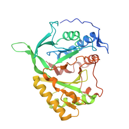
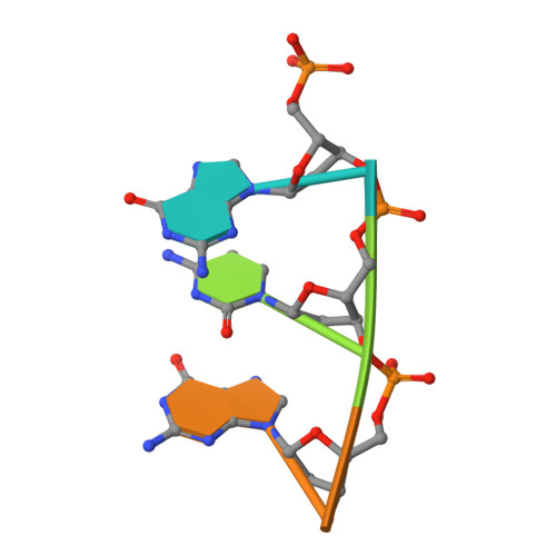
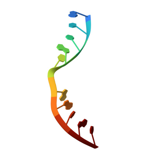
Molecular Basis for DNA Double-Strand Break Annealing and Primer Extension by an NHEJ DNA Polymerase.

Brissett, N.C.Martin, M.J.Bartlett, E.J.Bianchi, J.Blanco, L.Doherty, A.J.

(2013) Cell Rep 5: 1108-1120

  • DOI: https://doi.org/10.1016/j.celrep.2013.10.016
  • Primary Citation of Related Structures:  
    4MKY

  • PubMed Abstract: 

    Nonhomologous end-joining (NHEJ) is one of the major DNA double-strand break (DSB) repair pathways. The mechanisms by which breaks are competently brought together and extended during NHEJ is poorly understood. As polymerases extend DNA in a 5'-3' direction by nucleotide addition to a primer, it is unclear how NHEJ polymerases fill in break termini containing 3' overhangs that lack a primer strand. Here, we describe, at the molecular level, how prokaryotic NHEJ polymerases configure a primer-template substrate by annealing the 3' overhanging strands from opposing breaks, forming a gapped intermediate that can be extended in trans. We identify structural elements that facilitate docking of the 3' ends in the active sites of adjacent polymerases and reveal how the termini act as primers for extension of the annealed break, thus explaining how such DSBs are extended in trans. This study clarifies how polymerases couple break-synapsis to catalysis, providing a molecular mechanism to explain how primer extension is achieved on DNA breaks.


  • Organizational Affiliation

    Genome Damage and Stability Centre, University of Sussex, Brighton BN1 9RQ, UK.


Macromolecules

Find similar proteins by:  (by identity cutoff)  |  3D Structure
Entity ID: 1
MoleculeChains Sequence LengthOrganismDetailsImage
DNA ligase-like protein Rv0938/MT0965
A, B, C, D
303Mycobacterium tuberculosisMutation(s): 0 
Gene Names: MT0965MTCY08D9.01cMTCY10D7.36cRv0938
UniProt
Find proteins for P9WNV3 (Mycobacterium tuberculosis (strain ATCC 25618 / H37Rv))
Explore P9WNV3 
Go to UniProtKB:  P9WNV3
Entity Groups  
Sequence Clusters30% Identity50% Identity70% Identity90% Identity95% Identity100% Identity
UniProt GroupP9WNV3
Sequence Annotations
Expand
  • Reference Sequence

Find similar nucleic acids by:  Sequence   |   3D Structure  

Entity ID: 2
MoleculeChains LengthOrganismImage
5'-D(P*DGP*DCP*DGP*DGP*DC)-3'E,
F [auth G],
G [auth I],
H [auth K]
5N/A
Sequence Annotations
Expand
  • Reference Sequence

Find similar nucleic acids by:  Sequence   |   3D Structure  

Entity ID: 3
MoleculeChains LengthOrganismImage
5'-D(*DGP*DCP*DCP*DGP*DCP*DAP*DGP*DTP*DAP*DC)-3'I [auth F],
J [auth H],
K [auth J],
L
10N/A
Sequence Annotations
Expand
  • Reference Sequence
Experimental Data & Validation

Experimental Data

  • Method: X-RAY DIFFRACTION
  • Resolution: 2.40 Å
  • R-Value Free: 0.245 
  • R-Value Work: 0.197 
  • R-Value Observed: 0.200 
  • Space Group: P 1 21 1
Unit Cell:
Length ( Å )Angle ( ˚ )
a = 87.58α = 90
b = 80.11β = 111.62
c = 118.39γ = 90
Software Package:
Software NamePurpose
MOSFLMdata reduction
SCALAdata scaling
PHASERphasing
REFMACrefinement
PDB_EXTRACTdata extraction
CrystalCleardata collection

Structure Validation

View Full Validation Report


Entry History 

Deposition Data

Revision History  (Full details and data files)

  • Version 1.0: 2013-12-11
    Type: Initial release
  • Version 1.1: 2014-01-08
    Changes: Database references
  • Version 1.2: 2023-09-20
    Changes: Data collection, Database references, Refinement description