Structure based inhibitor design targeting glycogen phosphorylase b. Virtual screening, synthesis, biochemical and biological assessment of novel N-acyl-beta-d-glucopyranosylamines.
Parmenopoulou, V., Kantsadi, A.L., Tsirkone, V.G., Chatzileontiadou, D.S., Manta, S., Zographos, S.E., Molfeta, C., Archontis, G., Agius, L., Hayes, J.M., Leonidas, D.D., Komiotis, D.(2014) Bioorg Med Chem 22: 4810-4825
- PubMed: 25092521 Search on PubMed
- DOI: https://doi.org/10.1016/j.bmc.2014.06.058
- Primary Citation Related Structures:
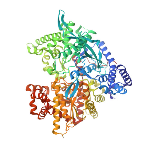
4MHO, 4MHS, 4MI3, 4MI6, 4MI9, 4MIC - PubMed Abstract:
Glycogen phosphorylase (GP) is a validated target for the development of new type 2 diabetes treatments. Exploiting the Zinc docking database, we report the in silico screening of 1888 N-acyl-β-d-glucopyranosylamines putative GP inhibitors differing only in their R groups. CombiGlide and GOLD docking programs with different scoring functions were employed with the best performing methods combined in a 'consensus scoring' approach to ranking of ligand binding affinities for the active site. Six selected candidates from the screening were then synthesized and their inhibitory potency was assessed both in vitro and ex vivo. Their inhibition constants' values, in vitro, ranged from 5 to 377μM while two of them were effective at causing inactivation of GP in rat hepatocytes at low μM concentrations. The crystal structures of GP in complex with the inhibitors were defined and provided the structural basis for their inhibitory potency and data for further structure based design of more potent inhibitors.
- Laboratory of Bio-Organic Chemistry, Department of Biochemistry and Biotechnology, University of Thessaly, 26 Ploutonos Str., 41221 Larissa, Greece.
Organizational Affiliation: