4M76 | pdb_00004m76

Integrin I domain of complement receptor 3 in complex with C3d


Experimental Data Snapshot

  • Method: X-RAY DIFFRACTION
  • Resolution: 2.80 Å
  • R-Value Free: 
    0.242 (Depositor), 0.247 (DCC) 
  • R-Value Work: 
    0.194 (Depositor), 0.199 (DCC) 
  • R-Value Observed: 
    0.198 (Depositor) 

Starting Models: experimental
View more details

wwPDB Validation 3D Report Full Report

Validation slider image for 4M76

This is version 1.5 of the entry. See complete history

Literature

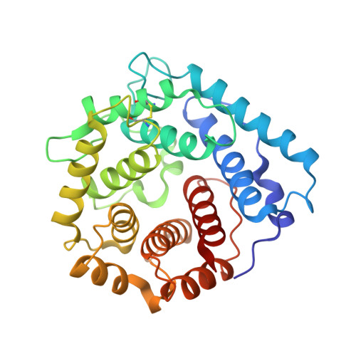
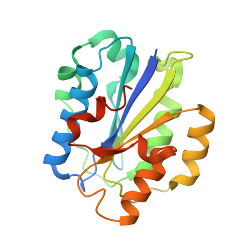
Structural insight on the recognition of surface-bound opsonins by the integrin I domain of complement receptor 3.

Bajic, G.Yatime, L.Sim, R.B.Vorup-Jensen, T.Andersen, G.R.

(2013) Proc Natl Acad Sci U S A 110: 16426-16431

  • DOI: https://doi.org/10.1073/pnas.1311261110
  • Primary Citation Related Structures: 
    4M76

  • PubMed Abstract: 

    Complement receptors (CRs), expressed notably on myeloid and lymphoid cells, play an essential function in the elimination of complement-opsonized pathogens and apoptotic/necrotic cells. In addition, these receptors are crucial for the cross-talk between the innate and adaptive branches of the immune system. CR3 (also known as Mac-1, integrin αMβ2, or CD11b/CD18) is expressed on all macrophages and recognizes iC3b on complement-opsonized objects, enabling their phagocytosis. We demonstrate that the C3d moiety of iC3b harbors the binding site for the CR3 αI domain, and our structure of the C3d:αI domain complex rationalizes the CR3 selectivity for iC3b. Based on extensive structural analysis, we suggest that the choice between a ligand glutamate or aspartate for coordination of a receptor metal ion-dependent adhesion site-bound metal ion is governed by the secondary structure of the ligand. Comparison of our structure to the CR2:C3d complex and the in vitro formation of a stable CR3:C3d:CR2 complex suggests a molecular mechanism for the hand-over of CR3-bound immune complexes from macrophages to CR2-presenting cells in lymph nodes.


  • Organizational Affiliation
    • Departments of Molecular Biology and Genetics and Biomedicine, Aarhus University, DK-8000 Aarhus, Denmark.

Macromolecule Content 

  • Total Structure Weight: 55.8 kDa 
  • Atom Count: 3,804 
  • Modeled Residue Count: 475 
  • Deposited Residue Count: 496 
  • Unique protein chains: 2

Macromolecules

Find similar proteins by:|  3D Structure
Entity ID: 1
MoleculeChains  Sequence LengthOrganismDetailsImage
Complement C3298Homo sapiensMutation(s): 1 
Gene Names: C3CPAMD1
UniProt & NIH Common Fund Data Resources
Find proteins for P01024 (Homo sapiens)
Explore P01024 
Go to UniProtKB:  P01024
PHAROS:  P01024
GTEx:  ENSG00000125730 
Entity Groups
Sequence Clusters30% Identity50% Identity70% Identity90% Identity95% Identity100% Identity
UniProt GroupP01024
Sequence Annotations
Expand
Reference Sequence
Find similar proteins by:|  3D Structure
Entity ID: 2
MoleculeChains  Sequence LengthOrganismDetailsImage
Integrin alpha-M198Homo sapiensMutation(s): 2 
Gene Names: CD11BCR3AITGAM
UniProt & NIH Common Fund Data Resources
Find proteins for P11215 (Homo sapiens)
Explore P11215 
Go to UniProtKB:  P11215
PHAROS:  P11215
GTEx:  ENSG00000169896 
Entity Groups
Sequence Clusters30% Identity50% Identity70% Identity90% Identity95% Identity100% Identity
UniProt GroupP11215
Sequence Annotations
Expand
Reference Sequence

Experimental Data & Validation

Experimental Data

  • Method: X-RAY DIFFRACTION
  • Resolution: 2.80 Å
  • R-Value Free:  0.242 (Depositor), 0.247 (DCC) 
  • R-Value Work:  0.194 (Depositor), 0.199 (DCC) 
  • R-Value Observed: 0.198 (Depositor) 
Space Group: C 1 2 1
Unit Cell:
Length ( Å )Angle ( ˚ )
a = 144.46α = 90
b = 65.12β = 115.69
c = 62.54γ = 90
Software Package:
Software NamePurpose
XSCALEdata scaling
PHASERphasing
PHENIXrefinement
PDB_EXTRACTdata extraction
XDSdata reduction

Structure Validation

View Full Validation Report


Entry History 

Deposition Data

Revision History  (Full details and data files)

  • Version 1.0: 2013-10-02
    Type: Initial release
  • Version 1.1: 2013-10-23
    Changes: Database references
  • Version 1.2: 2013-11-06
    Changes: Structure summary
  • Version 1.3: 2014-11-12
    Changes: Structure summary
  • Version 1.4: 2023-09-20
    Changes: Data collection, Database references, Derived calculations, Refinement description
  • Version 1.5: 2024-10-09
    Changes: Structure summary