Structural and Biochemical Analyses of Mycobacterium tuberculosis N-Acetylmuramyl-L-alanine Amidase Rv3717 Point to a Role in Peptidoglycan Fragment Recycling.
Prigozhin, D.M., Mavrici, D., Huizar, J.P., Vansell, H.J., Alber, T.(2013) J Biological Chem 288: 31549-31555
- PubMed: 24019530 Search on PubMedSearch on PubMed Central
- DOI: https://doi.org/10.1074/jbc.M113.510792
- Primary Citation Related Structures:
4M6G, 4M6H, 4M6I - PubMed Abstract:
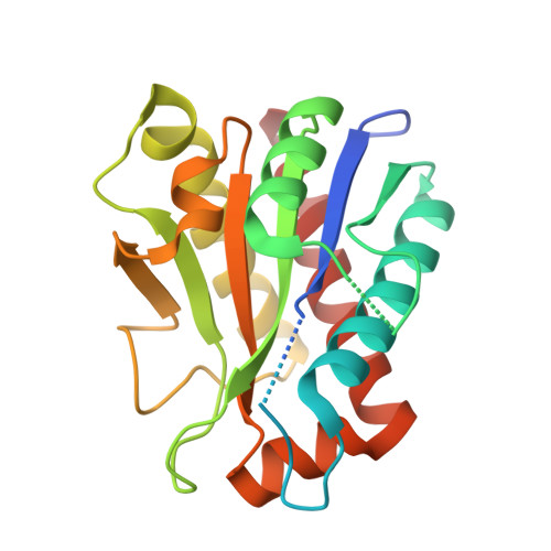
Peptidoglycan hydrolases are key enzymes in bacterial cell wall homeostasis. Understanding the substrate specificity and biochemical activity of peptidoglycan hydrolases in Mycobacterium tuberculosis is of special interest as it can aid in the development of new cell wall targeting therapeutics. In this study, we report biochemical and structural characterization of the mycobacterial N-acetylmuramyl-L-alanine amidase, Rv3717. The crystal structure of Rv3717 in complex with a dipeptide product shows that, compared with previously characterized peptidoglycan amidases, the enzyme contains an extra disulfide-bonded β-hairpin adjacent to the active site. The structure of two intermediates in assembly reveal that Zn(2+) binding rearranges active site residues, and disulfide formation promotes folding of the β-hairpin. Although Zn(2+) is required for hydrolysis of muramyl dipeptide, disulfide oxidation is not required for activity on this substrate. The orientation of the product in the active site suggests a role for a conserved glutamate (Glu-200) in catalysis; mutation of this residue abolishes activity. The product binds at the head of a closed tunnel, and the enzyme showed no activity on polymerized peptidoglycan. These results point to a potential role for Rv3717 in peptidoglycan fragment recycling.
- From the Department of Molecular and Cell Biology, University of California, Berkeley, California 94720-3220.
Organizational Affiliation: