4LRV

Crystal structure of DndE from Escherichia coli B7A involved in DNA phosphorothioation modification


Experimental Data Snapshot

  • Method: X-RAY DIFFRACTION
  • Resolution: 2.50 Å
  • R-Value Free: 0.242 
  • R-Value Work: 0.201 
  • R-Value Observed: 0.203 

wwPDB Validation   3D Report Full Report


This is version 1.1 of the entry. See complete history


Literature

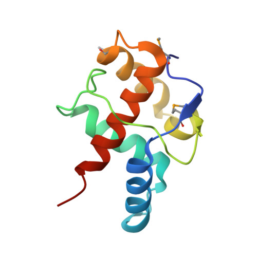
Structural insights into DndE from Escherichia coli B7A involved in DNA phosphorothioation modification

Hu, W.Wang, C.K.Liang, J.D.Zhang, T.L.Hu, Z.P.Wang, Z.J.Lan, W.X.Li, F.Wu, H.M.Ding, J.P.Wu, G.Deng, Z.X.Cao, C.

(2012) Cell Res 22: 1203-1206


Macromolecules
Find similar proteins by:  (by identity cutoff)  |  3D Structure
Entity ID: 1
MoleculeChains Sequence LengthOrganismDetailsImage
DNA sulfur modification protein DndE
A, B, C, D, E
A, B, C, D, E, F, G, H, I, J, K, L, M, N, O, P
109Escherichia coli B7AMutation(s): 0 
Gene Names: dndEEcB7A_5348
Entity Groups  
Sequence Clusters30% Identity50% Identity70% Identity90% Identity95% Identity100% Identity
Sequence Annotations
Expand
  • Reference Sequence
Small Molecules
Modified Residues  1 Unique
IDChains TypeFormula2D DiagramParent
MSE
Query on MSE
A, B, C, D, E
A, B, C, D, E, F, G, H, I, J, K, L, M, N, O, P
L-PEPTIDE LINKINGC5 H11 N O2 SeMET
Experimental Data & Validation

Experimental Data

  • Method: X-RAY DIFFRACTION
  • Resolution: 2.50 Å
  • R-Value Free: 0.242 
  • R-Value Work: 0.201 
  • R-Value Observed: 0.203 
  • Space Group: C 1 2 1
Unit Cell:
Length ( Å )Angle ( ˚ )
a = 97.845α = 90
b = 97.306β = 95.81
c = 218.532γ = 90
Software Package:
Software NamePurpose
HKL-2000data collection
PHENIXmodel building
REFMACrefinement
HKL-2000data reduction
HKL-2000data scaling
PHENIXphasing

Structure Validation

View Full Validation Report


Entry History 

Deposition Data

Revision History  (Full details and data files)

  • Version 1.0: 2013-08-28
    Type: Initial release
  • Version 1.1: 2013-09-04
    Changes: Experimental preparation