4LQ1 | pdb_00004lq1

Crystal Structure of E.Coli Branching Enzyme in complex with maltohexaose


Experimental Data Snapshot

  • Method: X-RAY DIFFRACTION
  • Resolution: 2.55 Å
  • R-Value Free: 
    0.224 (Depositor), 0.228 (DCC) 
  • R-Value Work: 
    0.178 (Depositor), 0.181 (DCC) 
  • R-Value Observed: 
    0.183 (Depositor) 

wwPDB Validation 3D Report Full Report

Validation slider image for 4LQ1

Ligand Structure Quality Assessment 


This is version 2.1 of the entry. See complete history

Literature

Crystal Structures of Escherichia coli Branching Enzyme in Complex with Linear Oligosaccharides.

Feng, L.Fawaz, R.Hovde, S.Gilbert, L.Chiou, J.Geiger, J.H.

(2015) Biochemistry 54: 6207-6218

  • DOI: https://doi.org/10.1021/acs.biochem.5b00228
  • Primary Citation Related Structures: 
    4LPC, 4LQ1

  • PubMed Abstract: 

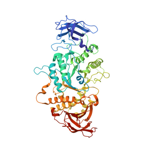
    Branching enzyme is responsible for all branching of glycogen and starch. It is an unusual member of the α-amylase family because it has both α-1,4-amylase activity and α-1,6-transferase activity [Drummond, G. S., et al. (1972) Eur. J. Biochem. 26, 168-176]. It also does not react with shorter glucans, though it will bind much longer substrates and substrate mimics [Binderup, K., et al. (2002) Arch. Biochem. Biophys. 397, 279-285]. In an effort to better understand how branching enzyme interacts with its polymeric substrate, we have determined the structure of Δ112 Escherichia coli branching enzyme bound to maltoheptaose and maltohexaose. Together, these structures define six distinct oligosaccharide binding sites on the surface of E. coli branching enzyme. Most of these binding sites surround the edge of the β-barrel domain and are quite far from the active site. Surprisingly, there is no evidence of oligosaccharide binding in the active site of the enzyme. The closest bound oligosaccharide resides almost 18 Å from the active site. Mutations to conserved residues in binding sites I and VI had a debilitating effect on the activity of the enzyme.


  • Organizational Affiliation
    • Department of Chemistry, Michigan State University , East Lansing, Michigan 48824, United States.

Macromolecule Content 

  • Total Structure Weight: 287.37 kDa 
  • Atom Count: 20,219 
  • Modeled Residue Count: 2,364 
  • Deposited Residue Count: 2,448 
  • Unique protein chains: 1

Macromolecules

Find similar proteins by:|  3D Structure
Entity ID: 1
MoleculeChains  Sequence LengthOrganismDetailsImage
1,4-alpha-glucan branching enzyme GlgB
A, B, C, D
612Escherichia coliMutation(s): 0 
Gene Names: glgBb3432JW3395
EC: 2.4.1.18
UniProt
Find proteins for P07762 (Escherichia coli (strain K12))
Explore P07762 
Go to UniProtKB:  P07762
Entity Groups
Sequence Clusters30% Identity50% Identity70% Identity90% Identity95% Identity100% Identity
UniProt GroupP07762
Sequence Annotations
Expand
Reference Sequence

Oligosaccharides

Help  
Entity ID: 2
MoleculeChains Length2D Diagram GlycosylationD Interactions
alpha-D-glucopyranose-(1-4)-alpha-D-glucopyranose
E, F, G, H, I
2N/A
Glycosylation Resources
GlyTouCan: G07411ON
GlyCosmos: G07411ON

Experimental Data & Validation

Experimental Data

  • Method: X-RAY DIFFRACTION
  • Resolution: 2.55 Å
  • R-Value Free:  0.224 (Depositor), 0.228 (DCC) 
  • R-Value Work:  0.178 (Depositor), 0.181 (DCC) 
  • R-Value Observed: 0.183 (Depositor) 
Space Group: P 1 21 1
Unit Cell:
Length ( Å )Angle ( ˚ )
a = 93.694α = 90
b = 104.113β = 91.85
c = 186.712γ = 90
Software Package:
Software NamePurpose
DENZOdata reduction
SCALEPACKdata scaling
MOLREPphasing
REFMACrefinement
PDB_EXTRACTdata extraction
HKL-2000data collection

Structure Validation

View Full Validation Report


Ligand Structure Quality Assessment 


Entry History 

Deposition Data

Revision History  (Full details and data files)

  • Version 1.0: 2015-04-01
    Type: Initial release
  • Version 1.1: 2015-09-23
    Changes: Database references
  • Version 1.2: 2016-04-06
    Changes: Database references
  • Version 1.3: 2017-11-15
    Changes: Refinement description
  • Version 2.0: 2020-07-29
    Type: Remediation
    Reason: Carbohydrate remediation
    Changes: Atomic model, Data collection, Derived calculations, Structure summary
  • Version 2.1: 2024-02-28
    Changes: Data collection, Database references, Structure summary