4LKT

Crystal Structure of Human Epidermal Fatty Acid Binding Protein (FABP5) in Complex with Linoleic Acid


Experimental Data Snapshot

  • Method: X-RAY DIFFRACTION
  • Resolution: 2.57 Å
  • R-Value Free: 0.259 
  • R-Value Work: 0.204 
  • R-Value Observed: 0.207 

wwPDB Validation   3D Report Full Report


Ligand Structure Quality Assessment 


This is version 1.3 of the entry. See complete history


Literature

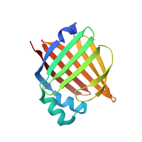
Structural basis for ligand regulation of the fatty acid-binding protein 5, peroxisome proliferator-activated receptor beta / delta (FABP5-PPAR beta / delta ) signaling pathway.

Armstrong, E.H.Goswami, D.Griffin, P.R.Noy, N.Ortlund, E.A.

(2014) J Biol Chem 289: 14941-14954

  • DOI: https://doi.org/10.1074/jbc.M113.514646
  • Primary Citation of Related Structures:  
    4LKP, 4LKT

  • PubMed Abstract: 

    Fatty acid-binding proteins (FABPs) are a widely expressed group of calycins that play a well established role in solubilizing cellular fatty acids. Recent studies, however, have recast FABPs as active participants in vital lipid-signaling pathways. FABP5, like its family members, displays a promiscuous ligand binding profile, capable of interacting with numerous long chain fatty acids of varying degrees of saturation. Certain "activating" fatty acids induce the protein's cytoplasmic to nuclear translocation, stimulating PPARβ/δ transactivation; however, the rules that govern this process remain unknown. Using a range of structural and biochemical techniques, we show that both linoleic and arachidonic acid elicit FABP5's translocation by permitting allosteric communication between the ligand-sensing β2 loop and a tertiary nuclear localization signal within the α-helical cap of the protein. Furthermore, we show that more saturated, nonactivating fatty acids inhibit nuclear localization signal formation by destabilizing this activation loop, thus implicating FABP5 specifically in cis-bonded, polyunsaturated fatty acid signaling.


  • Organizational Affiliation

    From the Department of Biochemistry, Discovery and Developmental Therapeutics, Winship Cancer Institute, Emory University School of Medicine, Atlanta, Georgia 30322.


Macromolecules
Find similar proteins by:  (by identity cutoff)  |  3D Structure
Entity ID: 1
MoleculeChains Sequence LengthOrganismDetailsImage
Fatty acid-binding protein, epidermal
A, B, C, D
138Homo sapiensMutation(s): 0 
Gene Names: FABP5
UniProt & NIH Common Fund Data Resources
Find proteins for Q01469 (Homo sapiens)
Explore Q01469 
Go to UniProtKB:  Q01469
PHAROS:  Q01469
GTEx:  ENSG00000164687 
Entity Groups  
Sequence Clusters30% Identity50% Identity70% Identity90% Identity95% Identity100% Identity
UniProt GroupQ01469
Sequence Annotations
Expand
  • Reference Sequence
Small Molecules
Ligands 7 Unique
IDChains Name / Formula / InChI Key2D Diagram3D Interactions
EIC
Query on EIC

Download Ideal Coordinates CCD File 
E [auth A],
K [auth B],
O [auth C],
V [auth D]
LINOLEIC ACID
C18 H32 O2
OYHQOLUKZRVURQ-HZJYTTRNSA-N
CIT
Query on CIT

Download Ideal Coordinates CCD File 
P [auth C]CITRIC ACID
C6 H8 O7
KRKNYBCHXYNGOX-UHFFFAOYSA-N
TAR
Query on TAR

Download Ideal Coordinates CCD File 
BA [auth D],
N [auth B],
U [auth C]
D(-)-TARTARIC ACID
C4 H6 O6
FEWJPZIEWOKRBE-LWMBPPNESA-N
SO4
Query on SO4

Download Ideal Coordinates CCD File 
AA [auth D],
J [auth A],
T [auth C]
SULFATE ION
O4 S
QAOWNCQODCNURD-UHFFFAOYSA-L
GOL
Query on GOL

Download Ideal Coordinates CCD File 
F [auth A]
G [auth A]
H [auth A]
I [auth A]
M [auth B]
F [auth A],
G [auth A],
H [auth A],
I [auth A],
M [auth B],
Q [auth C],
R [auth C],
W [auth D],
X [auth D]
GLYCEROL
C3 H8 O3
PEDCQBHIVMGVHV-UHFFFAOYSA-N
CL
Query on CL

Download Ideal Coordinates CCD File 
L [auth B]CHLORIDE ION
Cl
VEXZGXHMUGYJMC-UHFFFAOYSA-M
NH4
Query on NH4

Download Ideal Coordinates CCD File 
S [auth C],
Y [auth D],
Z [auth D]
AMMONIUM ION
H4 N
QGZKDVFQNNGYKY-UHFFFAOYSA-O
Binding Affinity Annotations 
IDSourceBinding Affinity
EIC BindingDB:  4LKT Ki: min: 1260, max: 3470 (nM) from 3 assay(s)
Experimental Data & Validation

Experimental Data

  • Method: X-RAY DIFFRACTION
  • Resolution: 2.57 Å
  • R-Value Free: 0.259 
  • R-Value Work: 0.204 
  • R-Value Observed: 0.207 
  • Space Group: P 32 2 1
Unit Cell:
Length ( Å )Angle ( ˚ )
a = 145.282α = 90
b = 145.282β = 90
c = 81.763γ = 120
Software Package:
Software NamePurpose
DENZOdata reduction
SCALEPACKdata scaling
PHASERphasing
PHENIXrefinement
PDB_EXTRACTdata extraction
HKL-2000data collection
HKL-2000data reduction
HKL-2000data scaling

Structure Validation

View Full Validation Report


Ligand Structure Quality Assessment 


Entry History 

Deposition Data

Revision History  (Full details and data files)

  • Version 1.0: 2014-03-26
    Type: Initial release
  • Version 1.1: 2016-04-13
    Changes: Database references
  • Version 1.2: 2017-11-15
    Changes: Refinement description
  • Version 1.3: 2024-02-28
    Changes: Data collection, Database references, Derived calculations