4L19 | pdb_00004l19

Matrix metalloproteinase-13 complexed with selective inhibitor compound Q1


Experimental Data Snapshot

  • Method: X-RAY DIFFRACTION
  • Resolution: 1.66 Å
  • R-Value Free: 
    0.199 (Depositor), 0.202 (DCC) 
  • R-Value Work: 
    0.134 (Depositor), 0.138 (DCC) 
  • R-Value Observed: 
    0.137 (Depositor) 

Starting Model: experimental
View more details

wwPDB Validation 3D Report Full Report

Validation slider image for 4L19

Ligand Structure Quality Assessment 


This is version 1.1 of the entry. See complete history

Literature

Characterization of selective exosite-binding inhibitors of matrix metalloproteinase 13 that prevent articular cartilage degradation in vitro.

Spicer, T.P.Jiang, J.Taylor, A.B.Choi, J.Y.Hart, P.J.Roush, W.R.Fields, G.B.Hodder, P.S.Minond, D.

(2014) J Med Chem 57: 9598-9611

  • DOI: https://doi.org/10.1021/jm501284e
  • Primary Citation Related Structures: 
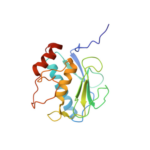
    4L19

  • PubMed Abstract: 

    Matrix metalloproteinase 13 (MMP-13) has been shown to be the main collagenase responsible for degradation of articular cartilage during osteoarthritis and therefore represents a target for drug development. As a result of high-throughput screening and structure-activity relationship studies, we identified a novel, highly selective class of MMP-13 inhibitors (compounds 1 (Q), 2 (Q1), and 3 (Q2)). Mechanistic characterization revealed a noncompetitive nature of these inhibitors with binding constants in the low micromolar range. Crystallographic analyses revealed two binding modes for compound 2 in the MMP-13 S1' subsite and in an S1/S2* subsite. Type II collagen- and cartilage-protective effects exhibited by compounds 1, 2, and 3 suggested that these compounds might be efficacious in future in vivo studies. Finally, these compounds were also highly selective when tested against a panel of 30 proteases, which, in combination with a good CYP inhibition profile, suggested low off-target toxicity and drug-drug interactions in humans.


  • Organizational Affiliation
    • Lead Identification Division, Translational Research Institute, ‡Department of Molecular Therapeutics, and §Department of Chemistry, Scripps Florida, The Scripps Research Institute , Jupiter, Florida 33458, United States.

Macromolecule Content 

  • Total Structure Weight: 40.28 kDa 
  • Atom Count: 3,061 
  • Modeled Residue Count: 336 
  • Deposited Residue Count: 342 
  • Unique protein chains: 1

Macromolecules

Find similar proteins by:|  3D Structure
Entity ID: 1
MoleculeChains  Sequence LengthOrganismDetailsImage
Collagenase 3
A, B
171Homo sapiensMutation(s): 0 
Gene Names: MMP13
EC: 3.4.24
UniProt & NIH Common Fund Data Resources
Find proteins for P45452 (Homo sapiens)
Explore P45452 
Go to UniProtKB:  P45452
PHAROS:  P45452
GTEx:  ENSG00000137745 
Entity Groups
Sequence Clusters30% Identity50% Identity70% Identity90% Identity95% Identity100% Identity
UniProt GroupP45452
Sequence Annotations
Expand
Reference Sequence

Small Molecules

Ligands 5 Unique
IDChains Name / Formula / InChI Key2D Diagram3D Interactions
1UA

Query on 1UA


Download:Ideal Coordinates CCD File
I [auth A],
J [auth A],
T [auth B]
2-[(4-methylbenzyl)sulfanyl]-3,5,6,7-tetrahydro-4H-cyclopenta[d]pyrimidin-4-one
C15 H16 N2 O S
SEZPRMPVJMVCBV-UHFFFAOYSA-N
GOL

Query on GOL


Download:Ideal Coordinates CCD File
K [auth A],
U [auth B],
V [auth B]
GLYCEROL
C3 H8 O3
PEDCQBHIVMGVHV-UHFFFAOYSA-N
ZN

Query on ZN


Download:Ideal Coordinates CCD File
C [auth A],
D [auth A],
E [auth A],
L [auth B],
M [auth B]
ZINC ION
Zn
PTFCDOFLOPIGGS-UHFFFAOYSA-N
FMT

Query on FMT


Download:Ideal Coordinates CCD File
H [auth A],
P [auth B],
Q [auth B],
R [auth B],
S [auth B]
FORMIC ACID
C H2 O2
BDAGIHXWWSANSR-UHFFFAOYSA-N
CA

Query on CA


Download:Ideal Coordinates CCD File
F [auth A],
G [auth A],
N [auth B],
O [auth B]
CALCIUM ION
Ca
BHPQYMZQTOCNFJ-UHFFFAOYSA-N
Binding Affinity Annotations 
IDSourceBinding Affinity
1UA BindingDB:  4L19 Ki: min: 800, max: 824 (nM) from 2 assay(s)
IC50: min: 400, max: 2400 (nM) from 2 assay(s)

Experimental Data & Validation

Experimental Data

  • Method: X-RAY DIFFRACTION
  • Resolution: 1.66 Å
  • R-Value Free:  0.199 (Depositor), 0.202 (DCC) 
  • R-Value Work:  0.134 (Depositor), 0.138 (DCC) 
  • R-Value Observed: 0.137 (Depositor) 
Space Group: C 1 2 1
Unit Cell:
Length ( Å )Angle ( ˚ )
a = 135.32α = 90
b = 36.029β = 130.23
c = 95.814γ = 90
Software Package:
Software NamePurpose
PHASERphasing
PHENIXrefinement
XDSdata reduction
XDSdata scaling

Structure Validation

View Full Validation Report


Ligand Structure Quality Assessment 


Entry History 

Deposition Data

Revision History  (Full details and data files)

  • Version 1.0: 2014-12-10
    Type: Initial release
  • Version 1.1: 2023-09-20
    Changes: Data collection, Database references, Derived calculations, Refinement description