4KMI

Crystal structure of 4-O-beta-D-mannosyl-D-glucose phosphorylase MGP complexed with PO4


Experimental Data Snapshot

  • Method: X-RAY DIFFRACTION
  • Resolution: 1.80 Å
  • R-Value Free: 0.233 
  • R-Value Work: 0.189 
  • R-Value Observed: 0.191 

wwPDB Validation   3D Report Full Report


This is version 1.2 of the entry. See complete history


Literature

Structure of Novel Enzyme in Mannan Biodegradation Process 4-O-beta-d-Mannosyl-d-Glucose Phosphorylase MGP

Nakae, S.Ito, S.Higa, M.Senoura, T.Wasaki, J.Hijikata, A.Shionyu, M.Ito, S.Shirai, T.

(2013) J Mol Biol 425: 4468-4478

  • DOI: https://doi.org/10.1016/j.jmb.2013.08.002
  • Primary Citation of Related Structures:  
    3WAS, 3WAT, 3WAU, 4KMI

  • PubMed Abstract: 

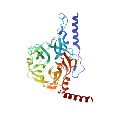
    The crystal structure of a novel component of the mannan biodegradation system, 4-O-β-D-mannosyl-D-glucose phosphorylase (MGP), was determined to a 1.68-Å resolution. The structure of the enzyme revealed a unique homohexameric structure, which was formed by using two helices attached to the N-terminus and C-terminus as a tab for sticking between subunits. The structures of MGP complexes with genuine substrates, 4-O-β-D-mannosyl-D-glucose and phosphate, and the product D-mannose-1-phosphate were also determined. The complex structures revealed that the invariant residue Asp131, which is supposed to be the general acid/base, did not exist close to the glycosidic Glc-O4 atom, which should be protonated in the catalytic reaction. Also, no solvent molecule that might mediate a proton transfer from Asp131 was observed in the substrate complex structure, suggesting that the catalytic mechanism of MGP is different from those of known disaccharide phosphorylases.


  • Organizational Affiliation

    Department of Bioscience, Nagahama Institute of Bio-science and Technology, and Japan Science and Technology Agency Institute for Bioinformatics Research and Development, 1266 Tamura, Nagahama 526-0829, Japan.


Macromolecules
Find similar proteins by:  (by identity cutoff)  |  3D Structure
Entity ID: 1
MoleculeChains Sequence LengthOrganismDetailsImage
4-O-beta-D-mannosyl-D-glucose phosphorylase
A, B
390Bacteroides fragilis NCTC 9343Mutation(s): 0 
Gene Names: BF0772
EC: 2.4.1.281
UniProt
Find proteins for Q5LH68 (Bacteroides fragilis (strain ATCC 25285 / DSM 2151 / CCUG 4856 / JCM 11019 / NCTC 9343 / Onslow))
Explore Q5LH68 
Go to UniProtKB:  Q5LH68
Entity Groups  
Sequence Clusters30% Identity50% Identity70% Identity90% Identity95% Identity100% Identity
UniProt GroupQ5LH68
Sequence Annotations
Expand
  • Reference Sequence
Experimental Data & Validation

Experimental Data

  • Method: X-RAY DIFFRACTION
  • Resolution: 1.80 Å
  • R-Value Free: 0.233 
  • R-Value Work: 0.189 
  • R-Value Observed: 0.191 
  • Space Group: H 3
Unit Cell:
Length ( Å )Angle ( ˚ )
a = 82.469α = 90
b = 82.469β = 90
c = 307.729γ = 120
Software Package:
Software NamePurpose
HKL-2000data collection
SOLVEphasing
PHENIXrefinement
HKL-2000data reduction
SCALEPACKdata scaling

Structure Validation

View Full Validation Report


Entry History 

Deposition Data

Revision History  (Full details and data files)

  • Version 1.0: 2013-09-04
    Type: Initial release
  • Version 1.1: 2013-11-20
    Changes: Database references
  • Version 1.2: 2023-11-08
    Changes: Data collection, Database references, Derived calculations, Refinement description