The putative propeptide of MycP1 in mycobacterial type VII secretion system does not inhibit protease activity but improves protein stability.
Sun, D., Liu, Q., He, Y., Wang, C., Wu, F., Tian, C., Zang, J.(2013) Protein Cell 4: 921-931
- PubMed: 24248472 Search on PubMedSearch on PubMed Central
- DOI: https://doi.org/10.1007/s13238-013-3089-7
- Primary Citation Related Structures:
4KB5, 4M1Z - PubMed Abstract:
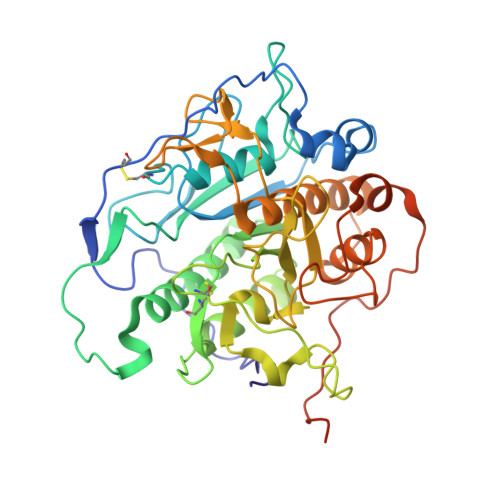
Mycosin-1 protease (MycP1) is a serine protease anchored to the inner membrane of Mycobacterium tuberculosis, and is essential in virulence factor secretion through the ESX-1 type VII secretion system (T7SS). Bacterial physiology studies demonstrated that MycP1 plays a dual role in the regulation of ESX-1 secretion and virulence, primarily through cleavage of its secretion substrate EspB. MycP1 contains a putative N-terminal inhibitory propeptide and a catalytic triad of Asp-His-Ser, classic hallmarks of a subtilase family serine protease. The MycP1 propeptide was previously reported to be initially inactive and activated after prolonged incubation. In this study, we have determined crystal structures of MycP1 with (MycP1²⁴⁻⁴²²) and without (MycP1⁶³⁻⁴²²) the propeptide, and conducted EspB cleavage assays using the two proteins. Very high structural similarity was observed in the two crystal structures. Interestingly, protease assays demonstrated positive EspB cleavage for both proteins, indicating that the putative propeptide does not inhibit protease activity. Molecular dynamic simulations showed higher rigidity in regions guarding the entrance to the catalytic site in MycP1²⁴⁻⁴²² than in MycP1⁶³⁻⁴²², suggesting that the putative propeptide might contribute to the conformational stability of the active site cleft and surrounding regions.
- National Laboratory for Physical Science at the Microscale and School of Life Sciences, University of Science and Technology of China, Hefei, 230026, China.
Organizational Affiliation: