Recognition of the thomsen-friedenreich pancarcinoma carbohydrate antigen by a lamprey variable lymphocyte receptor.
Luo, M., Velikovsky, C.A., Yang, X., Siddiqui, M.A., Hong, X., Barchi, J.J., Gildersleeve, J.C., Pancer, Z., Mariuzza, R.A.(2013) J Biological Chem 288: 23597-23606
- PubMed: 23782692 Search on PubMedSearch on PubMed Central
- DOI: https://doi.org/10.1074/jbc.M113.480467
- Primary Citation Related Structures:
4K5U, 4K79 - PubMed Abstract:
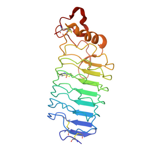
Variable lymphocyte receptors (VLRs) are leucine-rich repeat proteins that mediate adaptive immunity in jawless vertebrates. VLRs were recently shown to recognize glycans, such as the tumor-associated Thomsen-Friedenreich antigen (TFα; Galβ1-3GalNAcα), with a selectivity rivaling or exceeding that of lectins and antibodies. To understand the basis for TFα recognition by one such VLR (VLRB.aGPA.23), we measured thermodynamic parameters for the binding interaction and determined the structure of the VLRB.aGPA.23-TFα complex to 2.2 Å resolution. In the structure, four tryptophan residues form a tight hydrophobic cage encasing the TFα disaccharide that completely excludes buried water molecules. This cage together with hydrogen bonding of sugar hydroxyls to polar side chains explains the exquisite selectivity of VLRB.aGPA.23. The topology of the glycan-binding site of VLRB.aGPA.23 differs markedly from those of lectins or antibodies, which typically consist of long, convex grooves for accommodating the oligosaccharide. Instead, the TFα disaccharide is sandwiched between a variable loop and the concave surface of the VLR formed by the β-strands of the leucine-rich repeat modules. Longer oligosaccharides are predicted to extend perpendicularly across the β-strands, requiring them to bend to match the concavity of the VLR solenoid.
- University of Maryland Institute for Bioscience and Biotechnology Research, W. M. Keck Laboratory for Structural Biology, Rockville, Maryland 20850, USA.
Organizational Affiliation: