4JQG | pdb_00004jqg

Crystal structure of an inactive mutant of MMP-9 catalytic domain in complex with a fluorogenic synthetic peptidic substrate with a fluorine atom.


Experimental Data Snapshot

  • Method: X-RAY DIFFRACTION
  • Resolution: 1.85 Å
  • R-Value Free: 
    0.242 (Depositor), 0.256 (DCC) 
  • R-Value Work: 
    0.193 (Depositor) 
  • R-Value Observed: 
    0.196 (Depositor) 

Starting Model: experimental
View more details

wwPDB Validation 3D Report Full Report

Validation slider image for 4JQG

This is version 1.4 of the entry. See complete history

Literature

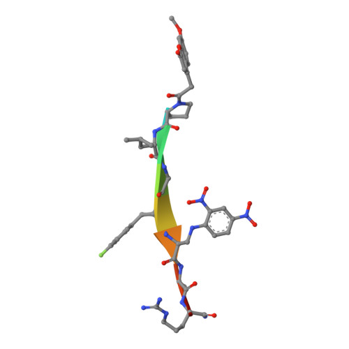
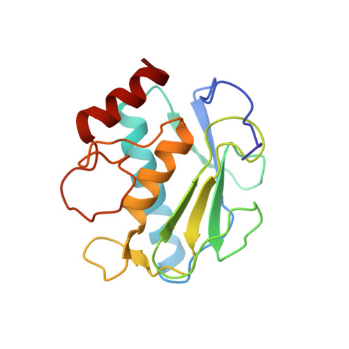
Halogen Bonding Controls Selectivity of FRET Substrate Probes for MMP-9.

Tranchant, I.Vera, L.Czarny, B.Amoura, M.Cassar, E.Beau, F.Stura, E.A.Dive, V.

(2014) Chem Biol 21: 408-413

  • DOI: https://doi.org/10.1016/j.chembiol.2014.01.008
  • Primary Citation Related Structures: 
    4JIJ, 4JQG

  • PubMed Abstract: 

    Matrix metalloproteinases (MMPs) are a large family of zinc-dependent endoproteases that catalyze cleavage of extracellular matrix and nonmatrix proteins. MMPs play a role in tissue remodeling, and their uncontrolled activity is associated with number of diseases, including tumor metastasis. Thus, there is a need to develop methods to monitor MMP activity, and number of probes has been previously described. The key problem many probes encounter is the issue of selectivity, since 23 human MMPs, despite playing different physiological roles, have structurally similar active sites. Here, we introduce the halogen bonding concept into the probe design and show that the probe containing iodine exhibits an unprecedented selectivity for MMP-9. We provide structure-based explanation for the selectivity, confirming that it is due to formation of the halogen bond that supports catalysis, and we highlight the value of exploring halogen bonding in the context of selective probe design.


  • Organizational Affiliation
    • CEA, iBiTec-S, Service d'Ingénierie Moléculaire des Protéines (SIMOPRO), Labex LERMIT, CE-Saclay, 91191 Gif sur Yvette Cedex, France.

Macromolecule Content 

  • Total Structure Weight: 40.33 kDa 
  • Atom Count: 3,322 
  • Modeled Residue Count: 346 
  • Deposited Residue Count: 346 
  • Unique protein chains: 2

Macromolecules

Find similar proteins by:  Sequence   |   3D Structure  
Entity ID: 1
MoleculeChains  Sequence LengthOrganismDetailsImage
fluorogenic peptidic substrate (8MC)PLG(PFF)(DNW)AR(NH2)A [auth P],
C [auth Q]
9N/AMutation(s): 0 
Find similar proteins by:|  3D Structure
Entity ID: 2
MoleculeChains  Sequence LengthOrganismDetailsImage
Matrix metalloproteinase-9B [auth A],
D [auth B]
164Homo sapiensMutation(s): 1 
Gene Names: CLG4BMMP9
EC: 3.4.24.35
UniProt & NIH Common Fund Data Resources
Find proteins for P14780 (Homo sapiens)
Explore P14780 
Go to UniProtKB:  P14780
PHAROS:  P14780
GTEx:  ENSG00000100985 
Entity Groups
Sequence Clusters30% Identity50% Identity70% Identity90% Identity95% Identity100% Identity
UniProt GroupP14780
Sequence Annotations
Expand
Reference Sequence

Small Molecules

Ligands 8 Unique
IDChains Name / Formula / InChI Key2D Diagram3D Interactions
GOL

Query on GOL


Download:Ideal Coordinates CCD File
L [auth A]GLYCEROL
C3 H8 O3
PEDCQBHIVMGVHV-UHFFFAOYSA-N
SR

Query on SR


Download:Ideal Coordinates CCD File
I [auth A],
V [auth B],
W [auth B]
STRONTIUM ION
Sr
PWYYWQHXAPXYMF-UHFFFAOYSA-N
PGO

Query on PGO


Download:Ideal Coordinates CCD File
Q [auth A],
X [auth B]
S-1,2-PROPANEDIOL
C3 H8 O2
DNIAPMSPPWPWGF-VKHMYHEASA-N
ZN

Query on ZN


Download:Ideal Coordinates CCD File
F [auth A],
G [auth A],
S [auth B],
T [auth B]
ZINC ION
Zn
PTFCDOFLOPIGGS-UHFFFAOYSA-N
EDO

Query on EDO


Download:Ideal Coordinates CCD File
K [auth A]
M [auth A]
N [auth A]
O [auth A]
P [auth A]
K [auth A],
M [auth A],
N [auth A],
O [auth A],
P [auth A],
Y [auth B],
Z [auth B]
1,2-ETHANEDIOL
C2 H6 O2
LYCAIKOWRPUZTN-UHFFFAOYSA-N
AZI

Query on AZI


Download:Ideal Coordinates CCD File
E [auth P],
R [auth Q]
AZIDE ION
N3
IVRMZWNICZWHMI-UHFFFAOYSA-N
CA

Query on CA


Download:Ideal Coordinates CCD File
H [auth A],
J [auth A],
U [auth B]
CALCIUM ION
Ca
BHPQYMZQTOCNFJ-UHFFFAOYSA-N
NA

Query on NA


Download:Ideal Coordinates CCD File
AA [auth B],
BA [auth B],
CA [auth B]
SODIUM ION
Na
FKNQFGJONOIPTF-UHFFFAOYSA-N
Modified Residues  2 Unique
IDChains TypeFormula2D DiagramParent
DNW
Query on DNW
A [auth P],
C [auth Q]
L-PEPTIDE LINKINGC9 H10 N4 O6ALA
PFF
Query on PFF
A [auth P],
C [auth Q]
L-PEPTIDE LINKINGC9 H10 F N O2PHE

Experimental Data & Validation

Experimental Data

  • Method: X-RAY DIFFRACTION
  • Resolution: 1.85 Å
  • R-Value Free:  0.242 (Depositor), 0.256 (DCC) 
  • R-Value Work:  0.193 (Depositor) 
  • R-Value Observed: 0.196 (Depositor) 
Space Group: P 21 21 21
Unit Cell:
Length ( Å )Angle ( ˚ )
a = 34.14α = 90
b = 57.48β = 90
c = 171.42γ = 90
Software Package:
Software NamePurpose
DNAdata collection
MOLREPphasing
PHENIXrefinement
XDSdata reduction
XDSdata scaling

Structure Validation

View Full Validation Report


Entry History 

Deposition Data

Revision History  (Full details and data files)

  • Version 1.0: 2014-03-12
    Type: Initial release
  • Version 1.1: 2014-04-16
    Changes: Database references
  • Version 1.2: 2017-08-16
    Changes: Data collection, Refinement description, Source and taxonomy
  • Version 1.3: 2023-09-20
    Changes: Data collection, Database references, Derived calculations, Refinement description
  • Version 1.4: 2023-12-06
    Changes: Data collection