4JDH | pdb_00004jdh

Crystal structure of Serine/threonine-protein kinase PAK 4 in complex with Paktide T peptide substrate


Experimental Data Snapshot

  • Method: X-RAY DIFFRACTION
  • Resolution: 2.00 Å
  • R-Value Free: 
    0.224 (Depositor), 0.225 (DCC) 
  • R-Value Work: 
    0.183 (Depositor), 0.182 (DCC) 
  • R-Value Observed: 
    0.185 (Depositor) 

Starting Model: experimental
View more details

wwPDB Validation 3D Report Full Report

Validation slider image for 4JDH

This is version 1.2 of the entry. See complete history

Literature

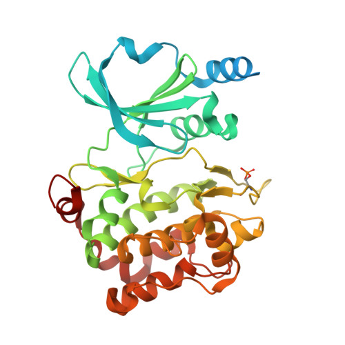
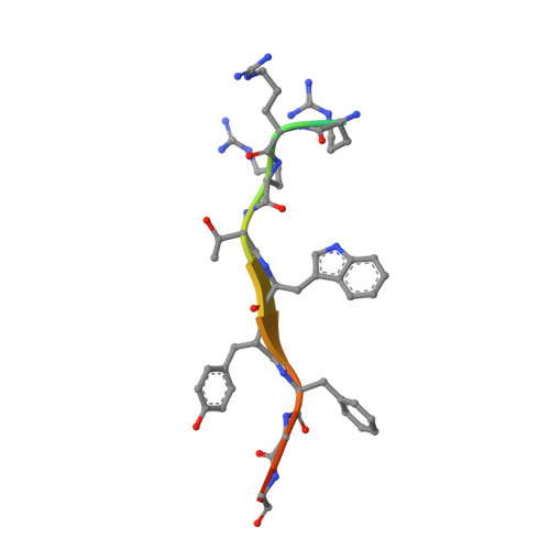
Identification of a major determinant for serine-threonine kinase phosphoacceptor specificity.

Chen, C.Ha, B.H.Thevenin, A.F.Lou, H.J.Zhang, R.Yip, K.Y.Peterson, J.R.Gerstein, M.Kim, P.M.Filippakopoulos, P.Knapp, S.Boggon, T.J.Turk, B.E.

(2014) Mol Cell 53: 140-147

  • DOI: https://doi.org/10.1016/j.molcel.2013.11.013
  • Primary Citation Related Structures: 
    4JDH, 4JDI, 4JDJ, 4JDK

  • PubMed Abstract: 

    Eukaryotic protein kinases are generally classified as being either tyrosine or serine-threonine specific. Though not evident from inspection of their primary sequences, many serine-threonine kinases display a significant preference for serine or threonine as the phosphoacceptor residue. Here we show that a residue located in the kinase activation segment, which we term the "DFG+1" residue, acts as a major determinant for serine-threonine phosphorylation site specificity. Mutation of this residue was sufficient to switch the phosphorylation site preference for multiple kinases, including the serine-specific kinase PAK4 and the threonine-specific kinase MST4. Kinetic analysis of peptide substrate phosphorylation and crystal structures of PAK4-peptide complexes suggested that phosphoacceptor residue preference is not mediated by stronger binding of the favored substrate. Rather, favored kinase-phosphoacceptor combinations likely promote a conformation optimal for catalysis. Understanding the rules governing kinase phosphoacceptor preference allows kinases to be classified as serine or threonine specific based on their sequence.


  • Organizational Affiliation
    • Department of Pharmacology, Yale University School of Medicine, New Haven, CT 06520, USA.

Macromolecule Content 

  • Total Structure Weight: 40.94 kDa 
  • Atom Count: 2,455 
  • Modeled Residue Count: 299 
  • Deposited Residue Count: 361 
  • Unique protein chains: 2

Macromolecules

Find similar proteins by:|  3D Structure
Entity ID: 1
MoleculeChains  Sequence LengthOrganismDetailsImage
Serine/threonine-protein kinase PAK 4346Homo sapiensMutation(s): 0 
Gene Names: KIAA1142PAK4
EC: 2.7.11.1
UniProt & NIH Common Fund Data Resources
Find proteins for O96013 (Homo sapiens)
Explore O96013 
Go to UniProtKB:  O96013
PHAROS:  O96013
GTEx:  ENSG00000130669 
Entity Groups
Sequence Clusters30% Identity50% Identity70% Identity90% Identity95% Identity100% Identity
UniProt GroupO96013
Sequence Annotations
Expand
Reference Sequence
Find similar proteins by:  Sequence   |   3D Structure  
Entity ID: 2
MoleculeChains  Sequence LengthOrganismDetailsImage
Paktide T15N/AMutation(s): 0 

Small Molecules

Modified Residues  1 Unique
IDChains TypeFormula2D DiagramParent
SEP
Query on SEP
A
L-PEPTIDE LINKINGC3 H8 N O6 PSER

Experimental Data & Validation

Experimental Data

  • Method: X-RAY DIFFRACTION
  • Resolution: 2.00 Å
  • R-Value Free:  0.224 (Depositor), 0.225 (DCC) 
  • R-Value Work:  0.183 (Depositor), 0.182 (DCC) 
  • R-Value Observed: 0.185 (Depositor) 
Space Group: P 41 21 2
Unit Cell:
Length ( Å )Angle ( ˚ )
a = 61.926α = 90
b = 61.926β = 90
c = 179.397γ = 90
Software Package:
Software NamePurpose
CBASSdata collection
PHASERphasing
REFMACrefinement
HKL-2000data reduction
HKL-2000data scaling

Structure Validation

View Full Validation Report


Entry History 

Deposition Data

Revision History  (Full details and data files)

  • Version 1.0: 2014-01-29
    Type: Initial release
  • Version 1.1: 2023-09-20
    Changes: Data collection, Database references, Derived calculations, Refinement description
  • Version 1.2: 2024-11-20
    Changes: Structure summary