4J2L

Crystal Structure of AXH domain complexed with Capicua


Experimental Data Snapshot

  • Method: X-RAY DIFFRACTION
  • Resolution: 3.15 Å
  • R-Value Free: 0.294 
  • R-Value Work: 0.236 
  • R-Value Observed: 0.236 

wwPDB Validation   3D Report Full Report


This is version 1.1 of the entry. See complete history


Literature

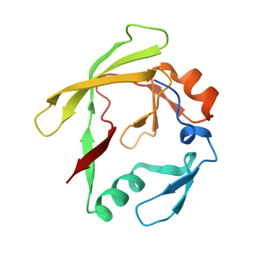
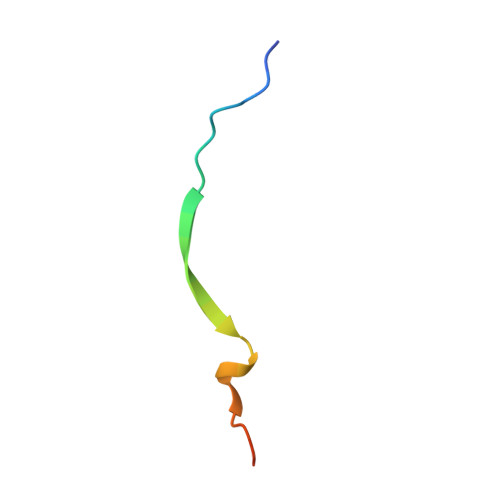
Structural basis of protein complex formation and reconfiguration by polyglutamine disease protein Ataxin-1 and Capicua

Kim, E.Lu, H.-C.Zoghbi, H.Y.Song, J.-J.

(2013) Genes Dev 27: 590-595

  • DOI: https://doi.org/10.1101/gad.212068.112
  • Primary Citation of Related Structures:  
    4J2J, 4J2L

  • PubMed Abstract: 

    Spinocerebellar ataxia type 1 (SCA1) is a dominantly inherited neurodegenerative disease caused by polyglutamine expansion in Ataxin-1 (ATXN1). ATXN1 binds to the transcriptional repressor Capicua (CIC), and the interaction plays a critical role in SCA1 pathogenesis whereby reducing CIC levels rescues SCA1-like phenotypes in a mouse model. The ATXN1/HBP1 (AXH) domain of ATXN1 mediates its homodimerization as well as the interaction with CIC. Here, we present the crystal structure of ATXN1's AXH domain bound to CIC and show that the binding pocket of the AXH domain to CIC overlaps with the homodimerization pocket of the AXH domain. Thus, the binding to CIC disrupts the homodimerization of ATXN1. Furthermore, the binding of CIC reconfigures the complex to allow another form of dimerization mediated by CIC, showing the intricacy of protein complex formation and reconfiguration by ATXN1 and CIC. Identifying the surfaces mediating the interactions between CIC and ATXN1 reveals a critical role for CIC in the reconfiguration of the AXH dimers and might provide insight into ways to target the ATXN1/CIC interactions to modulate SCA1 pathogenesis.


  • Organizational Affiliation

    Department of Biological Sciences, Graduate School of Nanoscience and Technology (World Class University), KAIST (Korea Advanced Institute of Science and Technology) Institute for the BioCentury, KAIST, Daejeon 305-701, Korea.


Macromolecules
Find similar proteins by:  (by identity cutoff)  |  3D Structure
Entity ID: 1
MoleculeChains Sequence LengthOrganismDetailsImage
Ataxin-1
A, B
129Homo sapiensMutation(s): 0 
Gene Names: ATXN1
UniProt & NIH Common Fund Data Resources
Find proteins for P54253 (Homo sapiens)
Explore P54253 
Go to UniProtKB:  P54253
PHAROS:  P54253
GTEx:  ENSG00000124788 
Entity Groups  
Sequence Clusters30% Identity50% Identity70% Identity90% Identity95% Identity100% Identity
UniProt GroupP54253
Sequence Annotations
Expand
  • Reference Sequence
Find similar proteins by:  (by identity cutoff)  |  3D Structure
Entity ID: 2
MoleculeChains Sequence LengthOrganismDetailsImage
Protein capicua homolog
C, D
28Homo sapiensMutation(s): 0 
Gene Names: CIC
UniProt & NIH Common Fund Data Resources
Find proteins for Q96RK0 (Homo sapiens)
Explore Q96RK0 
Go to UniProtKB:  Q96RK0
PHAROS:  Q96RK0
GTEx:  ENSG00000079432 
Entity Groups  
Sequence Clusters30% Identity50% Identity70% Identity90% Identity95% Identity100% Identity
UniProt GroupQ96RK0
Sequence Annotations
Expand
  • Reference Sequence
Experimental Data & Validation

Experimental Data

  • Method: X-RAY DIFFRACTION
  • Resolution: 3.15 Å
  • R-Value Free: 0.294 
  • R-Value Work: 0.236 
  • R-Value Observed: 0.236 
  • Space Group: P 43 2 2
Unit Cell:
Length ( Å )Angle ( ˚ )
a = 53.5α = 90
b = 53.5β = 90
c = 276.212γ = 90
Software Package:
Software NamePurpose
HKL-2000data collection
PHASERphasing
CNSrefinement
HKL-2000data reduction
HKL-2000data scaling

Structure Validation

View Full Validation Report


Entry History 

Deposition Data

Revision History  (Full details and data files)

  • Version 1.0: 2013-04-03
    Type: Initial release
  • Version 1.1: 2023-11-08
    Changes: Data collection, Database references, Refinement description